7ENL

MECHANISM OF ENOLASE: THE CRYSTAL STRUCTURE OF ENOLASE-MG2+-PHOSPHOGLYCERATE(SLASH) PHOSPHOENOLPYRUVATE COMPLEX AT 2.2-ANGSTROMS RESOLUTION


Experimental Data Snapshot

  • Method: X-RAY DIFFRACTION
  • Resolution: 2.20 Å
  • R-Value Observed: 0.169 

wwPDB Validation   3D Report Full Report


This is version 1.4 of the entry. See complete history


Literature

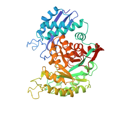
Mechanism of enolase: the crystal structure of enolase-Mg2(+)-2-phosphoglycerate/phosphoenolpyruvate complex at 2.2-A resolution.

Lebioda, L.Stec, B.

(1991) Biochemistry 30: 2817-2822

  • DOI: https://doi.org/10.1021/bi00225a012
  • Primary Citation of Related Structures:  
    7ENL

  • PubMed Abstract: 

    Enolase in the presence of Mg2+ catalyzes the elimination of H2O from 2-phosphoglyceric acid (PGA) to form phosphoenolpyruvate (PEP) and the reverse reaction, the hydration of PEP to PGA. The structure of the ternary complex yeast enolase-Mg2(+)-PGA/PEP has been determined by X-ray diffraction and refined by crystallographic restrained least-squares to an R = 16.9% for those data with I/sigma (I) greater than or equal to 2 to 2.2-A resolution with a good geometry of the model. The structure indicates the substrate molecule in the active site has its hydroxyl group coordinated to the Mg2+ ion. The carboxylic group interacts with the side chains of His373 and Lys396. The phosphate group is H-bonded to the guanidinium group of Arg374. A water molecule H-bonded to the carboxylic groups of Glu168 and Glu211 is located at a 2.6-A distance from carbon-2 of the substrate in the direction of its proton. We propose that this cluster functions as the base abstracting the proton in the catalytic process. The proton is probably transferred, first to the water molecule, then to Glu168, and further to the substrate hydroxyl to form a water molecule. Some analogy is apparent between the initial stages of the enolase reverse reaction, the hydration of PEP, and the proteolytic mechanism of the metallohydrolases carboxypeptidase A and thermolysin. The substrate/product binding is accompanied by large movements of loops Ser36-His43 and Ser158-Gly162. The role of these conformational changes is not clear at this time.


  • Organizational Affiliation

    Department of Chemistry, University of South Carolina, Columbia 29208.


Macromolecules
Find similar proteins by:  (by identity cutoff)  |  3D Structure
Entity ID: 1
MoleculeChains Sequence LengthOrganismDetailsImage
ENOLASE436Saccharomyces cerevisiaeMutation(s): 0 
EC: 4.2.1.11
UniProt
Find proteins for P00924 (Saccharomyces cerevisiae (strain ATCC 204508 / S288c))
Explore P00924 
Go to UniProtKB:  P00924
Entity Groups  
Sequence Clusters30% Identity50% Identity70% Identity90% Identity95% Identity100% Identity
UniProt GroupP00924
Sequence Annotations
Expand
  • Reference Sequence
Small Molecules
Ligands 2 Unique
IDChains Name / Formula / InChI Key2D Diagram3D Interactions
2PG
Query on 2PG

Download Ideal Coordinates CCD File 
C [auth A]2-PHOSPHOGLYCERIC ACID
C3 H7 O7 P
GXIURPTVHJPJLF-UWTATZPHSA-N
MG
Query on MG

Download Ideal Coordinates CCD File 
B [auth A]MAGNESIUM ION
Mg
JLVVSXFLKOJNIY-UHFFFAOYSA-N
Experimental Data & Validation

Experimental Data

  • Method: X-RAY DIFFRACTION
  • Resolution: 2.20 Å
  • R-Value Observed: 0.169 
  • Space Group: P 42 21 2
Unit Cell:
Length ( Å )Angle ( ˚ )
a = 122α = 90
b = 122β = 90
c = 67γ = 90
Software Package:
Software NamePurpose
PROLSQrefinement

Structure Validation

View Full Validation Report


Entry History 

Deposition Data

Revision History  (Full details and data files)

  • Version 1.0: 1992-04-15
    Type: Initial release
  • Version 1.1: 2008-03-03
    Changes: Version format compliance
  • Version 1.2: 2011-07-13
    Changes: Derived calculations, Version format compliance
  • Version 1.3: 2017-11-29
    Changes: Derived calculations, Other
  • Version 1.4: 2024-03-06
    Changes: Data collection, Database references, Derived calculations