


Crystal structure of apo streptavidin at cryogenic temperature
DeMirci, H.To be published.
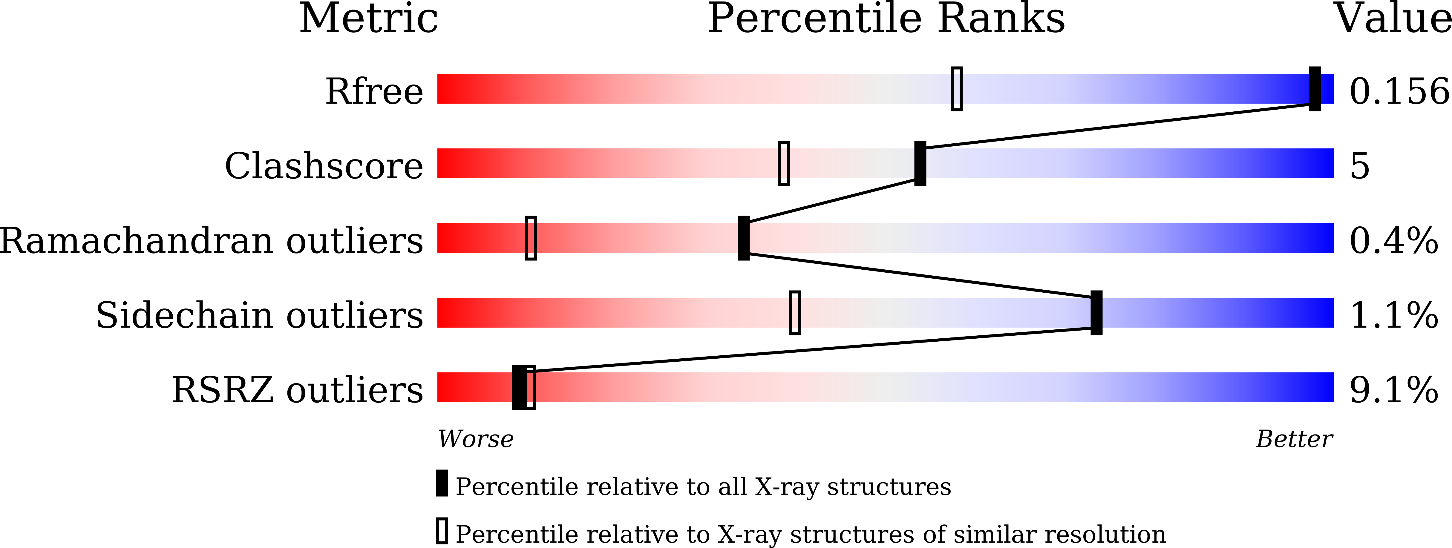
Experimental Data Snapshot
Entity ID: 1 | |||||
|---|---|---|---|---|---|
| Molecule | Chains | Sequence Length | Organism | Details | Image |
| Streptavidin | 123 | Streptomyces avidinii | Mutation(s): 0 |  | |
UniProt | |||||
Find proteins for P22629 (Streptomyces avidinii) Explore P22629 Go to UniProtKB: P22629 | |||||
Entity Groups | |||||
| Sequence Clusters | 30% Identity50% Identity70% Identity90% Identity95% Identity100% Identity | ||||
| UniProt Group | P22629 | ||||
Sequence AnnotationsExpand | |||||
| |||||
| Ligands 2 Unique | |||||
|---|---|---|---|---|---|
| ID | Chains | Name / Formula / InChI Key | 2D Diagram | 3D Interactions | |
| MPD (Subject of Investigation/LOI) Query on MPD | H [auth D] | (4S)-2-METHYL-2,4-PENTANEDIOL C6 H14 O2 SVTBMSDMJJWYQN-YFKPBYRVSA-N |  | ||
| MRD (Subject of Investigation/LOI) Query on MRD | E [auth A], F [auth C], G [auth D] | (4R)-2-METHYLPENTANE-2,4-DIOL C6 H14 O2 SVTBMSDMJJWYQN-RXMQYKEDSA-N |  | ||
| Length ( Å ) | Angle ( ˚ ) |
|---|---|
| a = 46.373 | α = 90 |
| b = 85.946 | β = 98.73 |
| c = 58.188 | γ = 90 |
| Software Name | Purpose |
|---|---|
| HKL-2000 | data scaling |
| PHENIX | refinement |
| PDB_EXTRACT | data extraction |
| XDS | data reduction |
| PHENIX | phasing |
| Funding Organization | Location | Grant Number |
|---|---|---|
| National Science Foundation (NSF, United States) | United States | NSF-1231306 |
| Other government | Turkey | 118C270 |
RCSB PDB (citation) is hosted by
RCSB PDB is a member of the

