7E96

Oxy-deoxy intermediate of 400 kDa giant hemoglobin at 69% oxygen saturation


Experimental Data Snapshot

  • Method: X-RAY DIFFRACTION
  • Resolution: 2.40 Å
  • R-Value Free: 0.238 
  • R-Value Work: 0.192 
  • R-Value Observed: 0.194 

wwPDB Validation   3D Report Full Report


Ligand Structure Quality Assessment 


This is version 1.2 of the entry. See complete history


Literature

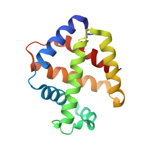
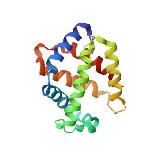
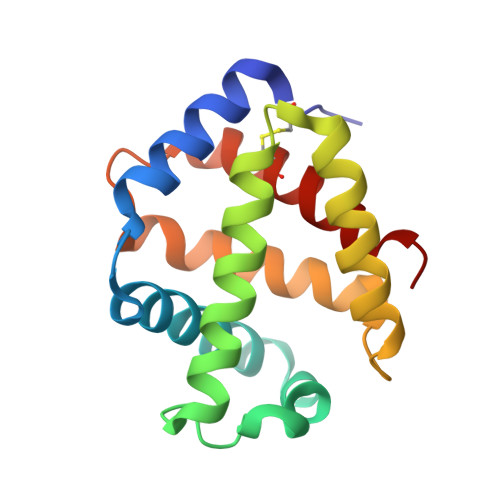
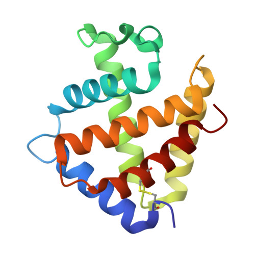
Coarse snapshots of oxygen-dissociation intermediates of a giant hemoglobin elucidated by determining the oxygen saturation in individual subunits in the crystalline state.

Numoto, N.Kawano, Y.Okumura, H.Baba, S.Fukumori, Y.Miki, K.Ito, N.

(2021) IUCrJ 8: 954-962

  • DOI: https://doi.org/10.1107/S2052252521009386
  • Primary Citation of Related Structures:  
    7E96, 7E97, 7E98, 7E99

  • PubMed Abstract: 

    Cooperative oxygen binding of hemoglobin (Hb) has been studied for over half a century as a representative example of the allostericity of proteins. The most important problem remaining to be solved is the lack of structural information on the intermediates between the oxygenated and deoxygenated forms. In order to characterize the intermediate structures, it is necessary to obtain intermediate-state crystals, determine their oxygen saturations and then determine the oxygen saturations of each of their constituent subunits, all of which are challenging issues even now. Here, intermediate forms of the 400 kDa giant Hb from the tubeworm Oligobrachia mashikoi are reported. To overcome the above problems without any artificial modifications to the protein or prosthetic groups, intermediate crystals of the giant Hb were prepared from fully oxygenated crystals by a soaking method. The oxygen saturation of the crystals was measured by in situ observation with a microspectrophotometer using thin plate crystals processed by an ultraviolet laser to avoid saturation of absorption. The oxygen saturation of each subunit was determined by occupancy refinement of the bound oxygen based on ambient temperature factors. The obtained structures reveal the detailed relationship between the structural transition and oxygen dissociation. The dimer subassembly of the giant Hb shows strong correlation with the local structural changes at the heme pockets. Although some local ternary-structural changes occur in the early stages of the structural transition, the associated global ternary-structural and quaternary-structural changes might arise at about 50% oxygen saturation. The models based on coarse snapshots of the allosteric transition support the conventional two-state model of Hbs and provide the missing pieces of the intermediate structures that are required for full understanding of the allosteric nature of Hbs in detail.


  • Organizational Affiliation

    Medical Research Institute, Tokyo Medical and Dental University (TMDU), 1-5-45 Yushima, Bunkyo-ku, Tokyo 113-8510, Japan.


Macromolecules
Find similar proteins by:  (by identity cutoff)  |  3D Structure
Entity ID: 1
MoleculeChains Sequence LengthOrganismDetailsImage
Extracellular giant hemoglobin major globin subunit A1140Oligobrachia mashikoiMutation(s): 0 
UniProt
Find proteins for Q7M419 (Oligobrachia mashikoi)
Explore Q7M419 
Go to UniProtKB:  Q7M419
Entity Groups  
Sequence Clusters30% Identity50% Identity70% Identity90% Identity95% Identity100% Identity
UniProt GroupQ7M419
Sequence Annotations
Expand
  • Reference Sequence
Find similar proteins by:  (by identity cutoff)  |  3D Structure
Entity ID: 2
MoleculeChains Sequence LengthOrganismDetailsImage
Extracellular giant hemoglobin major globin subunit A2142Oligobrachia mashikoiMutation(s): 0 
UniProt
Find proteins for Q7M413 (Oligobrachia mashikoi)
Explore Q7M413 
Go to UniProtKB:  Q7M413
Entity Groups  
Sequence Clusters30% Identity50% Identity70% Identity90% Identity95% Identity100% Identity
UniProt GroupQ7M413
Sequence Annotations
Expand
  • Reference Sequence
Find similar proteins by:  (by identity cutoff)  |  3D Structure
Entity ID: 3
MoleculeChains Sequence LengthOrganismDetailsImage
Extracellular giant hemoglobin major globin subunit B2147Oligobrachia mashikoiMutation(s): 0 
UniProt
Find proteins for Q7M418 (Oligobrachia mashikoi)
Explore Q7M418 
Go to UniProtKB:  Q7M418
Entity Groups  
Sequence Clusters30% Identity50% Identity70% Identity90% Identity95% Identity100% Identity
UniProt GroupQ7M418
Sequence Annotations
Expand
  • Reference Sequence
Find similar proteins by:  (by identity cutoff)  |  3D Structure
Entity ID: 4
MoleculeChains Sequence LengthOrganismDetailsImage
Giant hemoglobin B1b globin chain145Oligobrachia mashikoiMutation(s): 0 
UniProt
Find proteins for B1Q3G1 (Oligobrachia mashikoi)
Explore B1Q3G1 
Go to UniProtKB:  B1Q3G1
Entity Groups  
Sequence Clusters30% Identity50% Identity70% Identity90% Identity95% Identity100% Identity
UniProt GroupB1Q3G1
Sequence Annotations
Expand
  • Reference Sequence
Small Molecules
Ligands 3 Unique
IDChains Name / Formula / InChI Key2D Diagram3D Interactions
HEM (Subject of Investigation/LOI)
Query on HEM

Download Ideal Coordinates CCD File 
E [auth A],
H [auth B],
J [auth C],
L [auth D]
PROTOPORPHYRIN IX CONTAINING FE
C34 H32 Fe N4 O4
KABFMIBPWCXCRK-RGGAHWMASA-L
CA
Query on CA

Download Ideal Coordinates CCD File 
G [auth A]CALCIUM ION
Ca
BHPQYMZQTOCNFJ-UHFFFAOYSA-N
OXY (Subject of Investigation/LOI)
Query on OXY

Download Ideal Coordinates CCD File 
F [auth A],
I [auth B],
K [auth C],
M [auth D]
OXYGEN MOLECULE
O2
MYMOFIZGZYHOMD-UHFFFAOYSA-N
Experimental Data & Validation

Experimental Data

  • Method: X-RAY DIFFRACTION
  • Resolution: 2.40 Å
  • R-Value Free: 0.238 
  • R-Value Work: 0.192 
  • R-Value Observed: 0.194 
  • Space Group: H 3 2
Unit Cell:
Length ( Å )Angle ( ˚ )
a = 110.213α = 90
b = 110.213β = 90
c = 274.656γ = 120
Software Package:
Software NamePurpose
PHENIXrefinement
PHASERphasing
XDSdata processing
XDSdata reduction
XDSdata scaling

Structure Validation

View Full Validation Report


Ligand Structure Quality Assessment 


Entry History & Funding Information

Deposition Data


Funding OrganizationLocationGrant Number
Japan Society for the Promotion of Science (JSPS)Japan25870200
Japan Society for the Promotion of Science (JSPS)Japan15K20971

Revision History  (Full details and data files)

  • Version 1.0: 2021-10-06
    Type: Initial release
  • Version 1.1: 2022-02-16
    Changes: Database references
  • Version 1.2: 2023-11-29
    Changes: Data collection, Refinement description