7E6T

Structural insights into the activation of human calcium-sensing receptor


Experimental Data Snapshot

  • Method: ELECTRON MICROSCOPY
  • Resolution: 3.00 Å
  • Aggregation State: 2D ARRAY 
  • Reconstruction Method: SINGLE PARTICLE 

wwPDB Validation   3D Report Full Report


This is version 1.0 of the entry. See complete history


Literature

Structural insights into the activation of human calcium-sensing receptor.

Chen, X.Wang, L.Cui, Q.Ding, Z.Han, L.Kou, Y.Zhang, W.Wang, H.Jia, X.Dai, M.Shi, Z.Li, Y.Li, X.Geng, Y.

(2021) Elife 10

  • DOI: https://doi.org/10.7554/eLife.68578
  • Primary Citation of Related Structures:  
    7E6T, 7E6U

  • PubMed Abstract: 

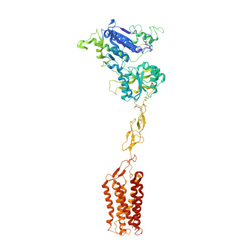
    Human calcium-sensing receptor (CaSR) is a G-protein-coupled receptor that maintains Ca 2+ homeostasis in serum. Here, we present the cryo-electron microscopy structures of the CaSR in the inactive and agonist+PAM bound states. Complemented with previously reported structures of CaSR, we show that in addition to the full inactive and active states, there are multiple intermediate states during the activation of CaSR. We used a negative allosteric nanobody to stabilize the CaSR in the fully inactive state and found a new binding site for Ca 2+ ion that acts as a composite agonist with L-amino acid to stabilize the closure of active Venus flytraps. Our data show that agonist binding leads to compaction of the dimer, proximity of the cysteine-rich domains, large-scale transitions of seven-transmembrane domains, and inter- and intrasubunit conformational changes of seven-transmembrane domains to accommodate downstream transducers. Our results reveal the structural basis for activation mechanisms of CaSR and clarify the mode of action of Ca 2+ ions and L-amino acid leading to the activation of the receptor.


  • Organizational Affiliation

    The CAS Key Laboratory of Receptor Research, Shanghai Institute of Materia Medica, Chinese Academy of Sciences, Shanghai, China.


Macromolecules
Find similar proteins by:  (by identity cutoff)  |  3D Structure
Entity ID: 1
MoleculeChains Sequence LengthOrganismDetailsImage
Extracellular calcium-sensing receptorA [auth B],
B [auth A]
862Homo sapiensMutation(s): 0 
Gene Names: CASRGPRC2APCAR1
Membrane Entity: Yes 
UniProt & NIH Common Fund Data Resources
Find proteins for P41180 (Homo sapiens)
Explore P41180 
Go to UniProtKB:  P41180
PHAROS:  P41180
GTEx:  ENSG00000036828 
Entity Groups  
Sequence Clusters30% Identity50% Identity70% Identity90% Identity95% Identity100% Identity
UniProt GroupP41180
Sequence Annotations
Expand
  • Reference Sequence
Small Molecules
Ligands 4 Unique
IDChains Name / Formula / InChI Key2D Diagram3D Interactions
NAG (Subject of Investigation/LOI)
Query on NAG

Download Ideal Coordinates CCD File 
G [auth B],
H [auth B],
N [auth A],
O [auth A]
2-acetamido-2-deoxy-beta-D-glucopyranose
C8 H15 N O6
OVRNDRQMDRJTHS-FMDGEEDCSA-N
TCR (Subject of Investigation/LOI)
Query on TCR

Download Ideal Coordinates CCD File 
I [auth B],
P [auth A]
CYCLOMETHYLTRYPTOPHAN
C12 H12 N2 O2
FSNCEEGOMTYXKY-JTQLQIEISA-N
PO4 (Subject of Investigation/LOI)
Query on PO4

Download Ideal Coordinates CCD File 
C [auth B],
J [auth A]
PHOSPHATE ION
O4 P
NBIIXXVUZAFLBC-UHFFFAOYSA-K
CA (Subject of Investigation/LOI)
Query on CA

Download Ideal Coordinates CCD File 
D [auth B]
E [auth B]
F [auth B]
K [auth A]
L [auth A]
D [auth B],
E [auth B],
F [auth B],
K [auth A],
L [auth A],
M [auth A]
CALCIUM ION
Ca
BHPQYMZQTOCNFJ-UHFFFAOYSA-N
Experimental Data & Validation

Experimental Data

  • Method: ELECTRON MICROSCOPY
  • Resolution: 3.00 Å
  • Aggregation State: 2D ARRAY 
  • Reconstruction Method: SINGLE PARTICLE 

Structure Validation

View Full Validation Report


Entry History & Funding Information

Deposition Data


Funding OrganizationLocationGrant Number
National Natural Science Foundation of China (NSFC)ChinaNo. 31670743
Chinese Academy of SciencesChina2015123456005

Revision History  (Full details and data files)

  • Version 1.0: 2021-09-22
    Type: Initial release