The hereditary mutation G51D unlocks a distinct fibril strain transmissible to wild-type alpha-synuclein.
Sun, Y., Long, H., Xia, W., Wang, K., Zhang, X., Sun, B., Cao, Q., Zhang, Y., Dai, B., Li, D., Liu, C.(2021) Nat Commun 12: 6252-6252
- PubMed: 34716315
- DOI: https://doi.org/10.1038/s41467-021-26433-2
- Primary Citation of Related Structures:
7E0F - PubMed Abstract:
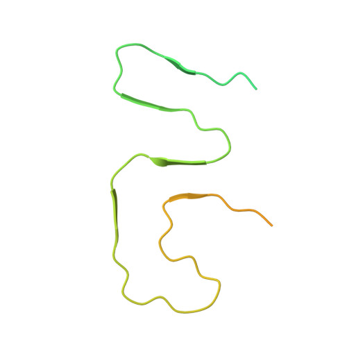
α-Synuclein (α-Syn) can form different fibril strains with distinct polymorphs and neuropathologies, which is associated with the clinicopathological variability in synucleinopathies. How different α-syn fibril strains are produced and selected under disease conditions remains poorly understood. In this study, we show that the hereditary mutation G51D induces α-syn to form a distinct fibril strain in vitro. The cryogenic electron microscopy (cryo-EM) structure of the G51D fibril strain was determined at 2.96 Å resolution. The G51D fibril displays a relatively small and extended serpentine fold distinct from other α-syn fibril structures. Moreover, we show by cryo-EM that wild-type (WT) α-syn can assembly into the G51D fibril strain via cross-seeding with G51D fibrils. Our study reveals a distinct structure of G51D fibril strain triggered by G51D mutation but feasibly adopted by both WT and G51D α-syn, which suggests the cross-seeding and strain selection of WT and mutant α-syn in familial Parkinson's disease (fPD).
Organizational Affiliation:
Interdisciplinary Research Center on Biology and Chemistry, Shanghai Institute of Organic Chemistry, Chinese Academy of Sciences, 201210, Shanghai, China.