


Crystal structure of a glutathione S-transferase SbGSTU6 from Salix babylonica in complex with glutathione
Zhuge, X.L., Yang, H.L.To be published.
Experimental Data Snapshot
wwPDB Validation 3D Report Full Report

Entity ID: 1 | |||||
|---|---|---|---|---|---|
| Molecule | Chains | Sequence Length | Organism | Details | Image |
| Glutathione S-transferase | 235 | Salix babylonica | Mutation(s): 1 Gene Names: GSTU6 | 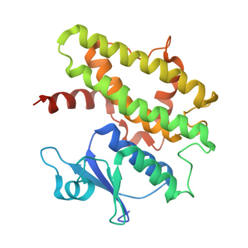 | |
UniProt | |||||
Find proteins for A0A4Y5R032 (Salix babylonica) Explore A0A4Y5R032 Go to UniProtKB: A0A4Y5R032 | |||||
Entity Groups | |||||
| Sequence Clusters | 30% Identity50% Identity70% Identity90% Identity95% Identity100% Identity | ||||
| UniProt Group | A0A4Y5R032 | ||||
Sequence AnnotationsExpand | |||||
| |||||
| Length ( Å ) | Angle ( ˚ ) |
|---|---|
| a = 104.882 | α = 90 |
| b = 104.882 | β = 90 |
| c = 53.09 | γ = 120 |
| Software Name | Purpose |
|---|---|
| PHENIX | refinement |
| HKL-2000 | data reduction |
| HKL-2000 | data scaling |
| PHASER | phasing |
| Funding Organization | Location | Grant Number |
|---|---|---|
| National Natural Science Foundation of China (NSFC) | China | NSFC 32071486 |
RCSB PDB (citation) is hosted by
RCSB PDB is a member of the

