7DOH

Crystal Structure of GD-26 Fab in Complex with TD Peptide from Haloarcula Marismortui Bacteriorhodopsin I


Experimental Data Snapshot

  • Method: X-RAY DIFFRACTION
  • Resolution: 1.45 Å
  • R-Value Free: 0.178 
  • R-Value Work: 0.130 
  • R-Value Observed: 0.133 

wwPDB Validation   3D Report Full Report


This is version 1.2 of the entry. See complete history


Literature

Structural basis of an epitope tagging system derived from Haloarcula marismortui bacteriorhodopsin I D94N and its monoclonal antibody GD-26.

Pao, P.J.Hsu, M.F.Chiang, M.H.Chen, C.T.Lee, C.C.Wang, A.H.

(2022) FEBS J 289: 730-747

  • DOI: https://doi.org/10.1111/febs.16184
  • Primary Citation of Related Structures:  
    7DOH

  • PubMed Abstract: 

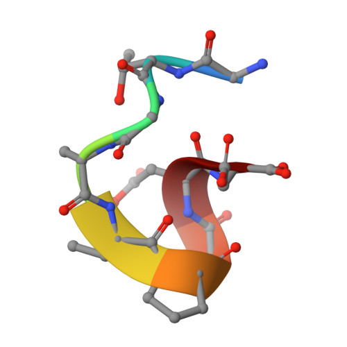
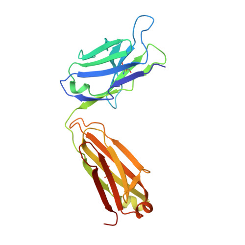
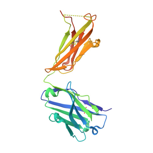
    Specific antibody interactions with short peptides have made epitope tagging systems a vital tool employed in virtually all fields of biological research. Here, we present a novel epitope tagging system comprised of a monoclonal antibody named GD-26, which recognises the TD peptide (GTGATPADD) derived from Haloarcula marismortui bacteriorhodopsin I (HmBRI) D94N mutant. The crystal structure of the antigen-binding fragment (Fab) of GD-26 complexed with the TD peptide was determined to a resolution of 1.45 Å. The TD peptide was found to adopt a 3 10 helix conformation within the binding cleft, providing a characteristic peptide structure for recognition by GD-26 Fab. Based on the structure information, polar and nonpolar forces collectively contribute to the strong binding. Attempts to engineer the TD peptide show that the proline residue is crucial for the formation of the 3 10 helix in order to fit into the binding cleft. Isothermal calorimetry (ITC) reported a dissociation constant K D of 12 ± 2.8 nm, indicating a strong interaction between the TD peptide and GD-26 Fab. High specificity of GD-26 IgG to the TD peptide was demonstrated by western blotting, ELISA and immunofluorescence as only TD-tagged proteins were detected, suggesting the effectiveness of the GD-26/TD peptide tagging system. In addition to already-existing epitope tags such as the FLAG tag and the ALFA tag adopting either extended or α-helix conformations, the unique 3 10 helix conformation of the TD peptide together with the corresponding monoclonal antibody GD-26 offers a novel tagging option for research.


  • Organizational Affiliation

    Institute of Biological Chemistry, Academia Sinica, Taipei, Taiwan.


Macromolecules

Find similar proteins by:  Sequence   |   3D Structure  

Entity ID: 1
MoleculeChains Sequence LengthOrganismDetailsImage
GLY-THR-GLY-ALA-THR-PRO-ALA-ASP-ASPA [auth I]9Haloarcula marismortuiMutation(s): 0 
UniProt
Find proteins for Q5UXY6 (Haloarcula marismortui (strain ATCC 43049 / DSM 3752 / JCM 8966 / VKM B-1809))
Explore Q5UXY6 
Go to UniProtKB:  Q5UXY6
Entity Groups  
UniProt GroupQ5UXY6
Sequence Annotations
Expand
  • Reference Sequence
Find similar proteins by:  (by identity cutoff)  |  3D Structure
Entity ID: 2
MoleculeChains Sequence LengthOrganismDetailsImage
GD-26 Fab L-chainB [auth L]219Mus musculusMutation(s): 0 
Entity Groups  
Sequence Clusters30% Identity50% Identity70% Identity90% Identity95% Identity100% Identity
Sequence Annotations
Expand
  • Reference Sequence
Find similar proteins by:  (by identity cutoff)  |  3D Structure
Entity ID: 3
MoleculeChains Sequence LengthOrganismDetailsImage
GD-26 Fab H-chainC [auth H]227Mus musculusMutation(s): 0 
Entity Groups  
Sequence Clusters30% Identity50% Identity70% Identity90% Identity95% Identity100% Identity
Sequence Annotations
Expand
  • Reference Sequence
Experimental Data & Validation

Experimental Data

  • Method: X-RAY DIFFRACTION
  • Resolution: 1.45 Å
  • R-Value Free: 0.178 
  • R-Value Work: 0.130 
  • R-Value Observed: 0.133 
  • Space Group: P 21 21 21
Unit Cell:
Length ( Å )Angle ( ˚ )
a = 51.427α = 90
b = 74.341β = 90
c = 108.103γ = 90
Software Package:
Software NamePurpose
PHENIXrefinement
HKL-3000data reduction
HKL-3000data scaling
PHENIXphasing

Structure Validation

View Full Validation Report


Entry History 

Deposition Data

Revision History  (Full details and data files)

  • Version 1.0: 2021-10-27
    Type: Initial release
  • Version 1.1: 2022-02-16
    Changes: Database references
  • Version 1.2: 2023-11-29
    Changes: Data collection, Derived calculations, Refinement description