7DO4

Crystal structure of CD97-CD55 complex

  • Classification: IMMUNE SYSTEM
  • Organism(s): Homo sapiens
  • Expression System: Homo sapiens
  • Mutation(s): No 

  • Deposited: 2020-12-11 Released: 2021-09-22 
  • Deposition Author(s): Niu, M., Song, G.
  • Funding Organization(s): National Natural Science Foundation of China (NSFC)

Experimental Data Snapshot

  • Method: X-RAY DIFFRACTION
  • Resolution: 3.20 Å
  • R-Value Free: 0.304 
  • R-Value Work: 0.267 
  • R-Value Observed: 0.271 

wwPDB Validation   3D Report Full Report


Ligand Structure Quality Assessment 


This is version 1.1 of the entry. See complete history


Literature

Structural basis for CD97 recognition of the decay-accelerating factor CD55 suggests mechanosensitive activation of adhesion GPCRs.

Niu, M.Xu, S.Yang, J.Yao, D.Li, N.Yan, J.Zhong, G.Song, G.

(2021) J Biol Chem 296: 100776-100776

  • DOI: https://doi.org/10.1016/j.jbc.2021.100776
  • Primary Citation of Related Structures:  
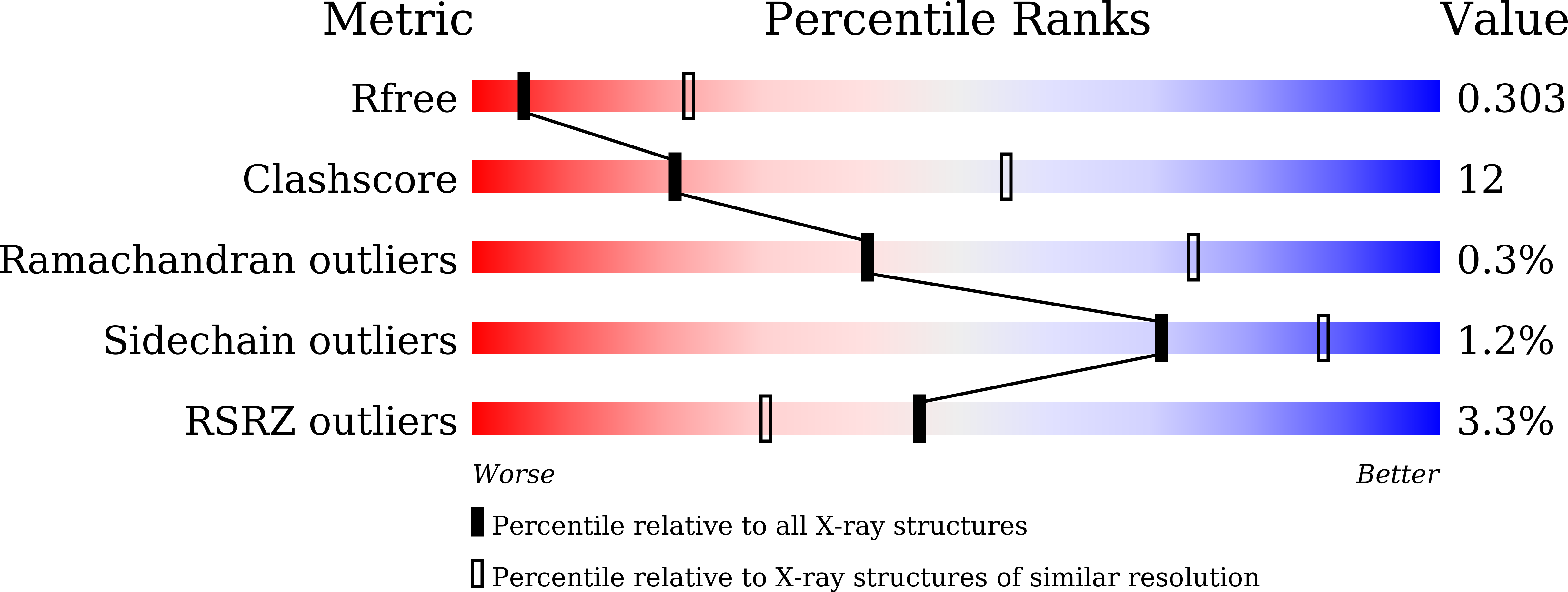
    7DO4

  • PubMed Abstract: 

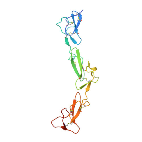
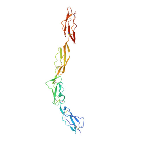
    The adhesion G protein-coupled receptor CD97 and its ligand complement decay-accelerating factor CD55 are important binding partners in the human immune system. Dysfunction in this binding has been linked to immune disorders such as multiple sclerosis and rheumatoid arthritis, as well as various cancers. Previous literatures have indicated that the CD97 includes 3 to 5 epidermal growth factor (EGF) domains at its N terminus and these EGF domains can bind to the N-terminal short consensus repeat (SCR) domains of CD55. However, the details of this interaction remain elusive, especially why the CD55 binds with the highest affinity to the shortest isoform of CD97 (EGF 1,2,5 ). Herein, we designed a chimeric expression construct with the EGF 1,2,5 domains of CD97 and the SCR 1-4 domains of CD55 connected by a flexible linker and determined the complex structure by crystallography. Our data reveal that the two proteins adopt an overall antiparallel binding mode involving the SCR 1-3 domains of CD55 and all three EGF domains of CD97. Mutagenesis data confirmed the importance of EGF 5 in the interaction and explained the binding specificity between CD55 and CD97. The architecture of CD55-CD97 binding mode together with kinetics suggests a force-resisting shearing stretch geometry when forces applied to the C termini of both proteins in the circulating environment. The potential of the CD55-CD97 complex to withstand tensile force may provide a basis for the mechanosensing mechanism for activation of adhesion G protein-coupled receptors.


  • Organizational Affiliation

    Shanghai Key Laboratory of Regulatory Biology, Institute of Biomedical Sciences and School of Life Sciences, East China Normal University, Shanghai, China.


Macromolecules
Find similar proteins by:  (by identity cutoff)  |  3D Structure
Entity ID: 1
MoleculeChains Sequence LengthOrganismDetailsImage
Isoform 2 of Adhesion G protein-coupled receptor E5145Homo sapiensMutation(s): 0 
Gene Names: ADGRE5CD97
UniProt & NIH Common Fund Data Resources
Find proteins for P48960 (Homo sapiens)
Explore P48960 
Go to UniProtKB:  P48960
PHAROS:  P48960
GTEx:  ENSG00000123146 
Entity Groups  
Sequence Clusters30% Identity50% Identity70% Identity90% Identity95% Identity100% Identity
UniProt GroupP48960
Sequence Annotations
Expand
  • Reference Sequence
Find similar proteins by:  (by identity cutoff)  |  3D Structure
Entity ID: 2
MoleculeChains Sequence LengthOrganismDetailsImage
Complement decay-accelerating factor251Homo sapiensMutation(s): 0 
Gene Names: CD55CRDAF
UniProt & NIH Common Fund Data Resources
Find proteins for P08174 (Homo sapiens)
Explore P08174 
Go to UniProtKB:  P08174
PHAROS:  P08174
GTEx:  ENSG00000196352 
Entity Groups  
Sequence Clusters30% Identity50% Identity70% Identity90% Identity95% Identity100% Identity
UniProt GroupP08174
Sequence Annotations
Expand
  • Reference Sequence
Oligosaccharides

Help

Entity ID: 3
MoleculeChains Length2D Diagram Glycosylation3D Interactions
2-acetamido-2-deoxy-beta-D-glucopyranose-(1-4)-2-acetamido-2-deoxy-beta-D-glucopyranose
C
2N-Glycosylation
Glycosylation Resources
GlyTouCan:  G42666HT
GlyCosmos:  G42666HT
GlyGen:  G42666HT
Experimental Data & Validation

Experimental Data

  • Method: X-RAY DIFFRACTION
  • Resolution: 3.20 Å
  • R-Value Free: 0.304 
  • R-Value Work: 0.267 
  • R-Value Observed: 0.271 
  • Space Group: P 1 21 1
Unit Cell:
Length ( Å )Angle ( ˚ )
a = 51.8α = 90
b = 44.25β = 98.7
c = 116.53γ = 90
Software Package:
Software NamePurpose
PHENIXrefinement
XDSdata reduction
Aimlessdata scaling
PHASERphasing
PDB_EXTRACTdata extraction

Structure Validation

View Full Validation Report


Ligand Structure Quality Assessment 


Entry History & Funding Information

Deposition Data


Funding OrganizationLocationGrant Number
National Natural Science Foundation of China (NSFC)China31770898

Revision History  (Full details and data files)

  • Version 1.0: 2021-09-22
    Type: Initial release
  • Version 1.1: 2023-11-29
    Changes: Data collection, Refinement description