7DNO

Characterization of Peptide Ligands Against WDR5 Isolated Using Phage Display Technique


Experimental Data Snapshot

  • Method: X-RAY DIFFRACTION
  • Resolution: 2.03 Å
  • R-Value Free: 0.227 
  • R-Value Work: 0.179 
  • R-Value Observed: 0.182 

wwPDB Validation   3D Report Full Report


This is version 1.2 of the entry. See complete history


Literature

Phage-Display Based Discovery and Characterization of Peptide Ligands against WDR5.

Cao, J.Fan, T.Li, Y.Du, Z.Chen, L.Wang, Y.Wang, X.Shen, J.Huang, X.Xiong, B.Cao, D.

(2021) Molecules 26

  • DOI: https://doi.org/10.3390/molecules26051225
  • Primary Citation of Related Structures:  
    7DNO

  • PubMed Abstract: 

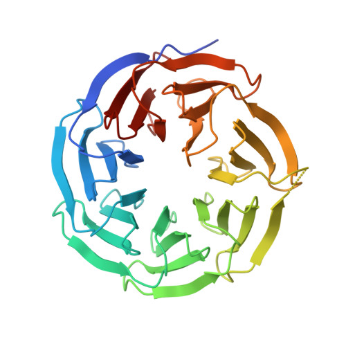
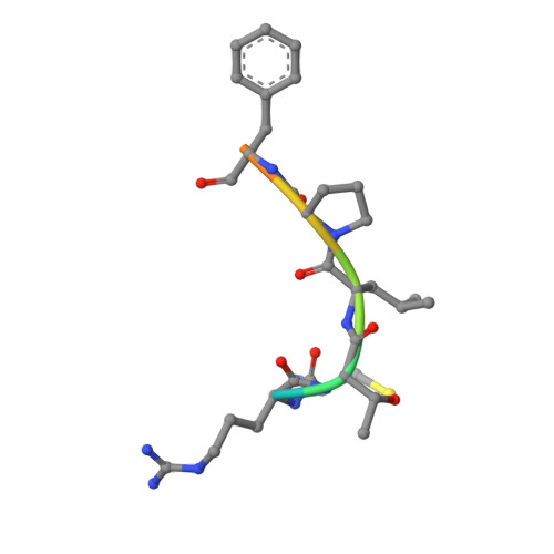
    WD40 is a ubiquitous domain presented in at least 361 human proteins and acts as scaffold to form protein complexes. Among them, WDR5 protein is an important mediator in several protein complexes to exert its functions in histone modification and chromatin remodeling. Therefore, it was considered as a promising epigenetic target involving in anti-cancer drug development. In view of the protein-protein interaction nature of WDR5, we initialized a campaign to discover new peptide-mimic inhibitors of WDR5. In current study, we utilized the phage display technique and screened with a disulfide-based cyclic peptide phage library. Five rounds of biopanning were performed and isolated clones were sequenced. By analyzing the sequences, total five peptides were synthesized for binding assay. The four peptides are shown to have the moderate binding affinity. Finally, the detailed binding interactions were revealed by solving a WDR5-peptide cocrystal structure.


  • Organizational Affiliation

    Department of College of Pharmacy, University of Chinese Academy of Sciences, No. 19A Yuquan Road, Beijing 100049, China.


Macromolecules
Find similar proteins by:  (by identity cutoff)  |  3D Structure
Entity ID: 1
MoleculeChains Sequence LengthOrganismDetailsImage
WD repeat-containing protein 5
A, B
313Homo sapiensMutation(s): 0 
Gene Names: WDR5BIG3
UniProt & NIH Common Fund Data Resources
Find proteins for P61964 (Homo sapiens)
Explore P61964 
Go to UniProtKB:  P61964
PHAROS:  P61964
GTEx:  ENSG00000196363 
Entity Groups  
Sequence Clusters30% Identity50% Identity70% Identity90% Identity95% Identity100% Identity
UniProt GroupP61964
Sequence Annotations
Expand
  • Reference Sequence

Find similar proteins by:  Sequence   |   3D Structure  

Entity ID: 2
MoleculeChains Sequence LengthOrganismDetailsImage
CYS-ARG-THR-LEU-PRO-PHE
C, D
9Homo sapiensMutation(s): 0 
Sequence Annotations
Expand
  • Reference Sequence
Experimental Data & Validation

Experimental Data

  • Method: X-RAY DIFFRACTION
  • Resolution: 2.03 Å
  • R-Value Free: 0.227 
  • R-Value Work: 0.179 
  • R-Value Observed: 0.182 
  • Space Group: P 1
Unit Cell:
Length ( Å )Angle ( ˚ )
a = 46.735α = 69.817
b = 53.912β = 88.79
c = 64.788γ = 74.163
Software Package:
Software NamePurpose
PHENIXrefinement
XDSdata reduction
XDSdata scaling
MOLREPphasing

Structure Validation

View Full Validation Report


Entry History 

Deposition Data

Revision History  (Full details and data files)

  • Version 1.0: 2021-02-10
    Type: Initial release
  • Version 1.1: 2022-02-23
    Changes: Database references, Structure summary
  • Version 1.2: 2023-11-29
    Changes: Data collection, Refinement description