7DNJ

K63-polyUb MDA5CARDs complex


Experimental Data Snapshot

  • Method: ELECTRON MICROSCOPY
  • Resolution: 3.30 Å
  • Aggregation State: PARTICLE 
  • Reconstruction Method: SINGLE PARTICLE 

wwPDB Validation   3D Report Full Report


This is version 1.1 of the entry. See complete history


Literature

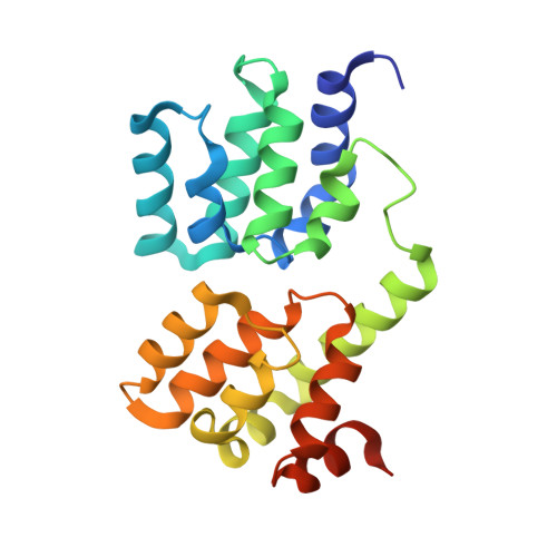
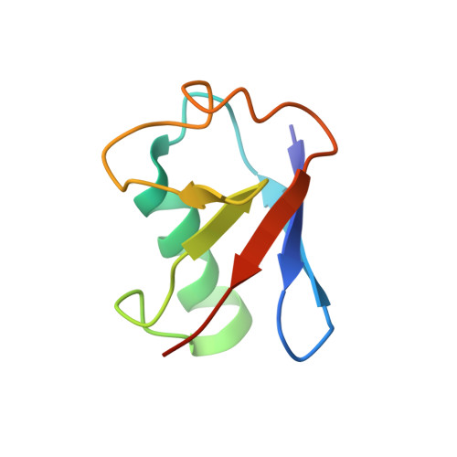
Ordered assembly of the cytosolic RNA-sensing MDA5-MAVS signaling complex via binding to unanchored K63-linked poly-ubiquitin chains.

Song, B.Chen, Y.Liu, X.Yuan, F.Tan, E.Y.J.Lei, Y.Song, N.Han, Y.Pascal, B.D.Griffin, P.R.Luo, C.Wu, B.Luo, D.Zheng, J.

(2021) Immunity 54: 2218

  • DOI: https://doi.org/10.1016/j.immuni.2021.09.008
  • Primary Citation of Related Structures:  
    7DNI, 7DNJ

  • PubMed Abstract: 

    The RNA sensor MDA5 recruits the signaling adaptor MAVS to initiate type I interferon signaling and downstream antiviral responses, a process that requires K63-linked polyubiquitin chains. Here, we examined the mechanisms whereby K63-polyUb chain regulate MDA5 activation. Only long unanchored K63-polyUb n (n ≥ 8) could mediate tetramerization of the caspase activation and recruitment domains of MDA5 ( MDA5 CARDs). Cryoelectron microscopy structures of a polyUb 13 -bound MDA5 CARDs tetramer and a polyUb 11 -bound MDA5 CARDs- MAVS CARD assembly revealed a tower-like formation, wherein eight Ubs tethered along the outer rim of the helical shell, bridging MDA5 CARDs and MAVS CARD tetramers into proximity. ATP binding and hydrolysis promoted the stabilization of RNA-bound MDA5 prior to MAVS activation via allosteric effects on CARDs-polyUb complex. Abundant ATP prevented basal activation of apo MDA5. Our findings reveal the ordered assembly of a MDA5 signaling complex competent to recruit and activate MAVS and highlight differences with RIG-I in terms of CARD orientation and Ub sensing that suggest different abilities to induce antiviral responses.


  • Organizational Affiliation

    Shanghai Institute of Materia Medica, Chinese Academy of Sciences, Shanghai 201203, China.


Macromolecules
Find similar proteins by:  (by identity cutoff)  |  3D Structure
Entity ID: 1
MoleculeChains Sequence LengthOrganismDetailsImage
Interferon-induced helicase C domain-containing protein 1
A, B, C, D
208Homo sapiensMutation(s): 0 
Gene Names: MDA5
EC: 3.6.4.13
UniProt & NIH Common Fund Data Resources
Find proteins for Q9BYX4 (Homo sapiens)
Explore Q9BYX4 
Go to UniProtKB:  Q9BYX4
PHAROS:  Q9BYX4
GTEx:  ENSG00000115267 
Entity Groups  
Sequence Clusters30% Identity50% Identity70% Identity90% Identity95% Identity100% Identity
UniProt GroupQ9BYX4
Sequence Annotations
Expand
  • Reference Sequence
Find similar proteins by:  (by identity cutoff)  |  3D Structure
Entity ID: 2
MoleculeChains Sequence LengthOrganismDetailsImage
Ubiquitin
E, F, G, H, I
E, F, G, H, I, J, K, L
76Homo sapiensMutation(s): 0 
Gene Names: UBB
UniProt & NIH Common Fund Data Resources
Find proteins for P0CG47 (Homo sapiens)
Explore P0CG47 
Go to UniProtKB:  P0CG47
PHAROS:  P0CG47
GTEx:  ENSG00000170315 
Entity Groups  
Sequence Clusters30% Identity50% Identity70% Identity90% Identity95% Identity100% Identity
UniProt GroupP0CG47
Sequence Annotations
Expand
  • Reference Sequence
Experimental Data & Validation

Experimental Data

  • Method: ELECTRON MICROSCOPY
  • Resolution: 3.30 Å
  • Aggregation State: PARTICLE 
  • Reconstruction Method: SINGLE PARTICLE 

Structure Validation

View Full Validation Report


Entry History & Funding Information

Deposition Data


Funding OrganizationLocationGrant Number
National Natural Science Foundation of China (NSFC)China81971538

Revision History  (Full details and data files)

  • Version 1.0: 2021-10-13
    Type: Initial release
  • Version 1.1: 2022-02-16
    Changes: Database references