7D8T

MITF bHLHLZ complex with M-box DNA


Experimental Data Snapshot

  • Method: X-RAY DIFFRACTION
  • Resolution: 3.20 Å
  • R-Value Free: 0.234 
  • R-Value Work: 0.204 
  • R-Value Observed: 0.206 

wwPDB Validation   3D Report Full Report


This is version 1.2 of the entry. See complete history


Literature

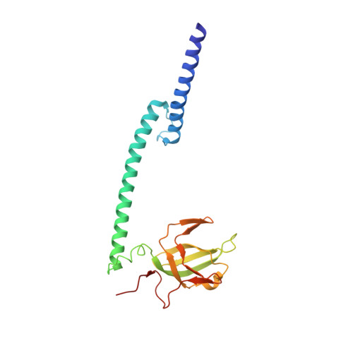
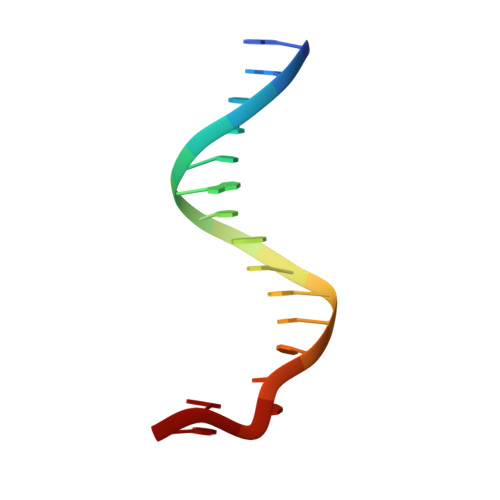
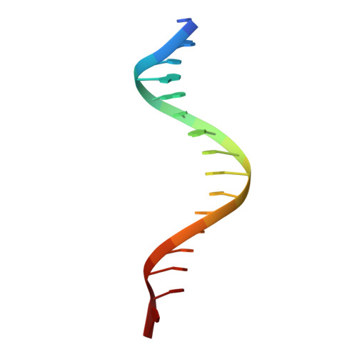
A unique hyperdynamic dimer interface permits small molecule perturbation of the melanoma oncoprotein MITF for melanoma therapy.

Liu, Z.Chen, K.Dai, J.Xu, P.Sun, W.Liu, W.Zhao, Z.Bennett, S.P.Li, P.Ma, T.Lin, Y.Kawakami, A.Yu, J.Wang, F.Wang, C.Li, M.Chase, P.Hodder, P.Spicer, T.P.Scampavia, L.Cao, C.Pan, L.Dong, J.Chen, Y.Yu, B.Guo, M.Fang, P.Fisher, D.E.Wang, J.

(2023) Cell Res 33: 55-70

  • DOI: https://doi.org/10.1038/s41422-022-00744-5
  • Primary Citation of Related Structures:  
    7D8R, 7D8S, 7D8T, 7EOD

  • PubMed Abstract: 

    Microphthalmia transcription factor (MITF) regulates melanocyte development and is the "lineage-specific survival" oncogene of melanoma. MITF is essential for melanoma initiation, progression, and relapse and has been considered an important therapeutic target; however, direct inhibition of MITF through small molecules is considered impossible, due to the absence of a ligand-binding pocket for drug design. Here, our structural analyses show that the structure of MITF is hyperdynamic because of its out-of-register leucine zipper with a 3-residue insertion. The dynamic MITF is highly vulnerable to dimer-disrupting mutations, as we observed that MITF loss-of-function mutations in human Waardenburg syndrome type 2 A are frequently located on the dimer interface and disrupt the dimer forming ability accordingly. These observations suggest a unique opportunity to inhibit MITF with small molecules capable of disrupting the MITF dimer. From a high throughput screening against 654,650 compounds, we discovered compound TT-012, which specifically binds to dynamic MITF and destroys the latter's dimer formation and DNA-binding ability. Using chromatin immunoprecipitation assay and RNA sequencing, we showed that TT-012 inhibits the transcriptional activity of MITF in B16F10 melanoma cells. In addition, TT-012 inhibits the growth of high-MITF melanoma cells, and inhibits the tumor growth and metastasis with tolerable toxicity to liver and immune cells in animal models. Together, this study demonstrates a unique hyperdynamic dimer interface in melanoma oncoprotein MITF, and reveals a novel approach to therapeutically suppress MITF activity.


  • Organizational Affiliation

    State Key Laboratory of Bioorganic and Natural Products Chemistry, Center for Excellence in Molecular Synthesis, Shanghai Institute of Organic Chemistry, University of Chinese Academy of Sciences, Chinese Academy of Sciences, Shanghai, China.


Macromolecules

Find similar proteins by:  (by identity cutoff)  |  3D Structure
Entity ID: 3
MoleculeChains Sequence LengthOrganismDetailsImage
Microphthalmia-associated transcription factor,Methionyl-tRNA synthetase beta subunitC [auth A],
D [auth B]
199Homo sapiensAquifex aeolicus VF5
This entity is chimeric
Mutation(s): 1 
Gene Names: MITFBHLHE32metG'aq_422
UniProt & NIH Common Fund Data Resources
Find proteins for O66738 (Aquifex aeolicus (strain VF5))
Explore O66738 
Go to UniProtKB:  O66738
Find proteins for O75030 (Homo sapiens)
Explore O75030 
Go to UniProtKB:  O75030
PHAROS:  O75030
GTEx:  ENSG00000187098 
Entity Groups  
Sequence Clusters30% Identity50% Identity70% Identity90% Identity95% Identity100% Identity
UniProt GroupsO66738O75030
Sequence Annotations
Expand
  • Reference Sequence

Find similar nucleic acids by:  Sequence   |   3D Structure  

Entity ID: 1
MoleculeChains LengthOrganismImage
DNA (5'-D(P*GP*GP*GP*AP*CP*AP*CP*AP*TP*GP*TP*TP*AP*CP*AP*G)-3')A [auth C]16Homo sapiens
Sequence Annotations
Expand
  • Reference Sequence

Find similar nucleic acids by:  Sequence   |   3D Structure  

Entity ID: 2
MoleculeChains LengthOrganismImage
DNA (5'-D(*TP*GP*TP*AP*AP*CP*AP*TP*GP*TP*GP*TP*CP*CP*CP*C)-3')B [auth D]16Homo sapiens
Sequence Annotations
Expand
  • Reference Sequence
Experimental Data & Validation

Experimental Data

  • Method: X-RAY DIFFRACTION
  • Resolution: 3.20 Å
  • R-Value Free: 0.234 
  • R-Value Work: 0.204 
  • R-Value Observed: 0.206 
  • Space Group: H 3 2
Unit Cell:
Length ( Å )Angle ( ˚ )
a = 207.26α = 90
b = 207.26β = 90
c = 163.16γ = 120
Software Package:
Software NamePurpose
PHENIXrefinement
SCALAdata scaling
PDB_EXTRACTdata extraction
HKL-2000data reduction
MOLREPphasing

Structure Validation

View Full Validation Report


Entry History & Funding Information

Deposition Data


Funding OrganizationLocationGrant Number
National Natural Science Foundation of China (NSFC)China21778064
National Natural Science Foundation of China (NSFC)China21778067
National Natural Science Foundation of China (NSFC)China21977108
National Natural Science Foundation of China (NSFC)China21977107

Revision History  (Full details and data files)

  • Version 1.0: 2021-10-13
    Type: Initial release
  • Version 1.1: 2023-09-27
    Changes: Data collection, Database references
  • Version 1.2: 2023-11-29
    Changes: Refinement description