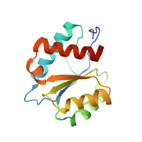
7D7U

Crystal structure of Ago2 MID domain in complex with 8-Br-adenosin-5'-monophosphate


Experimental Data Snapshot

  • Method: X-RAY DIFFRACTION
  • Resolution: 2.00 Å
  • R-Value Free: 0.247 
  • R-Value Work: 0.204 
  • R-Value Observed: 0.207 

wwPDB Validation   3D Report Full Report


Ligand Structure Quality Assessment 


This is version 1.2 of the entry. See complete history


Literature

siRNA potency enhancement via chemical modifications of nucleotide bases at the 5'-end of the siRNA guide strand.

Shinohara, F.Oashi, T.Harumoto, T.Nishikawa, T.Takayama, Y.Miyagi, H.Takahashi, Y.Nakajima, T.Sawada, T.Koda, Y.Makino, A.Sato, A.Hamaguchi, K.Suzuki, M.Yamamoto, J.Tomari, Y.Saito, J.I.

(2021) RNA 27: 163-173

  • DOI: https://doi.org/10.1261/rna.073783.119
  • Primary Citation of Related Structures:  
    7C6B, 7D7U

  • PubMed Abstract: 

    Small interfering RNAs (siRNAs) can be utilized not only as functional biological research tools but also as therapeutic agents. For the clinical use of siRNA as drugs, various chemical modifications have been used to improve the activity of siRNA drugs, and further chemical modifications are expected to improve the utility of siRNA therapeutics. As the 5' nucleobase of the guide strand affects the interaction between an siRNA and AGO2 and target cleavage activity, structural optimization of this specific position may be a useful strategy for improving siRNA activity. Here, using the in silico model of the complex between human AGO2 MID domain and nucleoside monophosphates, we screened and synthesized an original adenine-derived analog, 6-(3-(2-carboxyethyl)phenyl)purine (6-mCEPh-purine), that fits better than the natural nucleotide bases into the MID domain of AGO2. Introduction of the 6-mCEPh-purine analog at the 5'-end of the siRNA guide strand significantly enhanced target knockdown activity in both cultured cell lines and in vivo animal models. Our findings can help expand strategies for rationally optimizing siRNA activity via chemical modifications of nucleotide bases.


  • Organizational Affiliation

    Research Function Unit, R&D Division, Kyowa Kirin Co. Ltd., Chiyoda-ku, Tokyo 100-0004, Japan.


Macromolecules
Find similar proteins by:  (by identity cutoff)  |  3D Structure
Entity ID: 1
MoleculeChains Sequence LengthOrganismDetailsImage
Protein argonaute-2
A, B, C
139Homo sapiensMutation(s): 0 
Gene Names: AGO2EIF2C2
EC: 3.1.26
UniProt & NIH Common Fund Data Resources
Find proteins for Q9UKV8 (Homo sapiens)
Explore Q9UKV8 
Go to UniProtKB:  Q9UKV8
PHAROS:  Q9UKV8
GTEx:  ENSG00000123908 
Entity Groups  
Sequence Clusters30% Identity50% Identity70% Identity90% Identity95% Identity100% Identity
UniProt GroupQ9UKV8
Sequence Annotations
Expand
  • Reference Sequence
Small Molecules
Binding Affinity Annotations 
IDSourceBinding Affinity
8BR Binding MOAD:  7D7U Kd: 1.00e+5 (nM) from 1 assay(s)
Experimental Data & Validation

Experimental Data

  • Method: X-RAY DIFFRACTION
  • Resolution: 2.00 Å
  • R-Value Free: 0.247 
  • R-Value Work: 0.204 
  • R-Value Observed: 0.207 
  • Space Group: P 1
Unit Cell:
Length ( Å )Angle ( ˚ )
a = 47.17α = 105.83
b = 65.77β = 96.46
c = 40.79γ = 87.56
Software Package:
Software NamePurpose
MOSFLMdata reduction
Aimlessdata scaling
MOLREPphasing
REFMACrefinement
PDB_EXTRACTdata extraction

Structure Validation

View Full Validation Report


Ligand Structure Quality Assessment 


Entry History 

Deposition Data

Revision History  (Full details and data files)

  • Version 1.0: 2020-11-25
    Type: Initial release
  • Version 1.1: 2021-02-03
    Changes: Database references
  • Version 1.2: 2023-11-29
    Changes: Data collection, Database references, Refinement description