7D3S

Human SECR in complex with an engineered Gs heterotrimer


Experimental Data Snapshot

  • Method: ELECTRON MICROSCOPY
  • Resolution: 2.90 Å
  • Aggregation State: PARTICLE 
  • Reconstruction Method: SINGLE PARTICLE 

wwPDB Validation   3D Report Full Report


This is version 1.1 of the entry. See complete history


Literature

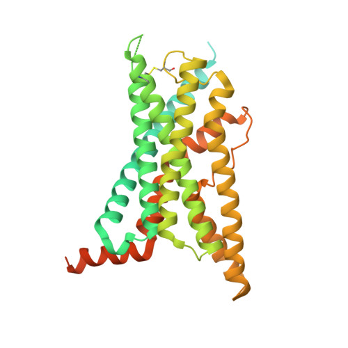
Structure of the human secretin receptor coupled to an engineered heterotrimeric G protein.

Fukuhara, S.Kobayashi, K.Kusakizako, T.Iida, W.Kato, M.Shihoya, W.Nureki, O.

(2020) Biochem Biophys Res Commun 533: 861-866

  • DOI: https://doi.org/10.1016/j.bbrc.2020.08.042
  • Primary Citation of Related Structures:  
    7D3S

  • PubMed Abstract: 

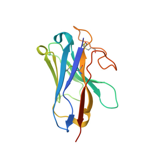
    Secretin is a gastrointestinal hormone that exerts multiple physiological functions via activation of the secretin receptor (SECR). SECR belongs to the class B G-protein-coupled receptors and is involved in various processes, such as regulation of the pH of the duodenal content, food intake, and water homeostasis. Here, we report a cryo-electron microscopy structure of human SECR bound to secretin and an engineered Gs heterotrimer. The structure revealed the basic architecture of SECR and the secretin binding mode. A structural comparison of the SECR and PAC1R transmembrane domains revealed that transmembrane helices 1 and 2 play a prominent role in secretin recognition. Moreover, the extracellular domain of SECR is perpendicular to the TMD, unlike that of PAC1R. This comparison revealed the diverged peptide recognition mechanisms of these receptors, which belong to the same subgroup. Our structural information will facilitate drug discovery research for clinical applications.


  • Organizational Affiliation

    Department of Biological Sciences, Graduate School of Science, The University of Tokyo, 7-3-1 Hongo, Bunkyo-ku, Tokyo, 113-0033, Japan; Department of Family Medicine, Graduate School of Medical and Dental Sciences, Tokyo Medical and Dental University, 1-5-45 Yushima, Bunkyo-ku, Tokyo, 113-8519, Japan.


Macromolecules
Find similar proteins by:  (by identity cutoff)  |  3D Structure
Entity ID: 1
MoleculeChains Sequence LengthOrganismDetailsImage
SecretinA [auth P]27Homo sapiensMutation(s): 0 
Membrane Entity: Yes 
UniProt & NIH Common Fund Data Resources
Find proteins for P09683 (Homo sapiens)
Explore P09683 
Go to UniProtKB:  P09683
PHAROS:  P09683
GTEx:  ENSG00000070031 
Entity Groups  
Sequence Clusters30% Identity50% Identity70% Identity90% Identity95% Identity100% Identity
UniProt GroupP09683
Sequence Annotations
Expand
  • Reference Sequence
Find similar proteins by:  (by identity cutoff)  |  3D Structure
Entity ID: 2
MoleculeChains Sequence LengthOrganismDetailsImage
Secretin receptorB [auth R]434Homo sapiensMutation(s): 0 
Gene Names: SCTR
Membrane Entity: Yes 
UniProt & NIH Common Fund Data Resources
Find proteins for P47872 (Homo sapiens)
Explore P47872 
Go to UniProtKB:  P47872
PHAROS:  P47872
GTEx:  ENSG00000080293 
Entity Groups  
Sequence Clusters30% Identity50% Identity70% Identity90% Identity95% Identity100% Identity
UniProt GroupP47872
Sequence Annotations
Expand
  • Reference Sequence
Find similar proteins by:  (by identity cutoff)  |  3D Structure
Entity ID: 3
MoleculeChains Sequence LengthOrganismDetailsImage
Guanine nucleotide-binding protein G(s) subunit alpha isoforms shortC [auth A]380Homo sapiensMutation(s): 7 
Gene Names: GNASGNAS1GSP
Membrane Entity: Yes 
UniProt & NIH Common Fund Data Resources
Find proteins for P63092 (Homo sapiens)
Explore P63092 
Go to UniProtKB:  P63092
PHAROS:  P63092
GTEx:  ENSG00000087460 
Entity Groups  
Sequence Clusters30% Identity50% Identity70% Identity90% Identity95% Identity100% Identity
UniProt GroupP63092
Sequence Annotations
Expand
  • Reference Sequence
Find similar proteins by:  (by identity cutoff)  |  3D Structure
Entity ID: 4
MoleculeChains Sequence LengthOrganismDetailsImage
Guanine nucleotide-binding protein G(I)/G(S)/G(T) subunit beta-1D [auth B]351Rattus norvegicusMutation(s): 0 
Gene Names: Gnb1
UniProt
Find proteins for P54311 (Rattus norvegicus)
Explore P54311 
Go to UniProtKB:  P54311
Entity Groups  
Sequence Clusters30% Identity50% Identity70% Identity90% Identity95% Identity100% Identity
UniProt GroupP54311
Sequence Annotations
Expand
  • Reference Sequence
Find similar proteins by:  (by identity cutoff)  |  3D Structure
Entity ID: 5
MoleculeChains Sequence LengthOrganismDetailsImage
Guanine nucleotide-binding protein G(I)/G(S)/G(O) subunit gamma-2E [auth G]68Bos taurusMutation(s): 0 
Gene Names: GNG2
Membrane Entity: Yes 
UniProt
Find proteins for P63212 (Bos taurus)
Explore P63212 
Go to UniProtKB:  P63212
Entity Groups  
Sequence Clusters30% Identity50% Identity70% Identity90% Identity95% Identity100% Identity
UniProt GroupP63212
Sequence Annotations
Expand
  • Reference Sequence
Find similar proteins by:  (by identity cutoff)  |  3D Structure
Entity ID: 6
MoleculeChains Sequence LengthOrganismDetailsImage
nanobody Nb35F [auth N]137unidentifiedMutation(s): 0 
Entity Groups  
Sequence Clusters30% Identity50% Identity70% Identity90% Identity95% Identity100% Identity
Sequence Annotations
Expand
  • Reference Sequence
Experimental Data & Validation

Experimental Data

  • Method: ELECTRON MICROSCOPY
  • Resolution: 2.90 Å
  • Aggregation State: PARTICLE 
  • Reconstruction Method: SINGLE PARTICLE 
EM Software:
TaskSoftware PackageVersion
MODEL REFINEMENTPHENIX1.14

Structure Validation

View Full Validation Report


Entry History 

Deposition Data

Revision History  (Full details and data files)

  • Version 1.0: 2020-11-04
    Type: Initial release
  • Version 1.1: 2020-12-30
    Changes: Database references