7CYM

Crystal structure of LI-Cadherin EC1-4


Experimental Data Snapshot

  • Method: X-RAY DIFFRACTION
  • Resolution: 2.70 Å
  • R-Value Free: 0.274 
  • R-Value Work: 0.222 
  • R-Value Observed: 0.225 

wwPDB Validation   3D Report Full Report


This is version 1.2 of the entry. See complete history


Literature

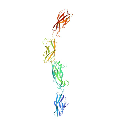
Mechanism of dimerization and structural features of human LI-cadherin.

Yui, A.Caaveiro, J.M.M.Kuroda, D.Nakakido, M.Nagatoishi, S.Goda, S.Maruno, T.Uchiyama, S.Tsumoto, K.

(2021) J Biol Chem 297: 101054-101054

  • DOI: https://doi.org/10.1016/j.jbc.2021.101054
  • Primary Citation of Related Structures:  
    7CYM, 7EV1

  • PubMed Abstract: 

    Liver intestine (LI)-cadherin is a member of the cadherin superfamily, which encompasses a group of Ca 2+ -dependent cell-adhesion proteins. The expression of LI-cadherin is observed on various types of cells in the human body, such as normal small intestine and colon cells, and gastric cancer cells. Because its expression is not observed on normal gastric cells, LI-cadherin is a promising target for gastric cancer imaging. However, because the cell adhesion mechanism of LI-cadherin has remained unknown, rational design of therapeutic molecules targeting this cadherin has been hampered. Here, we have studied the homodimerization mechanism of LI-cadherin. We report the crystal structure of the LI-cadherin homodimer containing its first four extracellular cadherin repeats (EC1-4). The EC1-4 homodimer exhibited a unique architecture different from that of other cadherins reported so far, driven by the interactions between EC2 of one protein chain and EC4 of the second protein chain. The crystal structure also revealed that LI-cadherin possesses a noncanonical calcium ion-free linker between the EC2 and EC3 domains. Various biochemical techniques and molecular dynamics simulations were employed to elucidate the mechanism of homodimerization. We also showed that the formation of the homodimer observed in the crystal structure is necessary for LI-cadherin-dependent cell adhesion by performing cell aggregation assays. Taken together, our data provide structural insights necessary to advance the use of LI-cadherin as a target for imaging gastric cancer.


  • Organizational Affiliation

    Department of Bioengineering, School of Engineering, The University of Tokyo, Tokyo, Japan.


Macromolecules
Find similar proteins by:  (by identity cutoff)  |  3D Structure
Entity ID: 1
MoleculeChains Sequence LengthOrganismDetailsImage
Cadherin-17
A, B
440Homo sapiensMutation(s): 0 
Gene Names: CDH17
UniProt & NIH Common Fund Data Resources
Find proteins for Q12864 (Homo sapiens)
Explore Q12864 
Go to UniProtKB:  Q12864
PHAROS:  Q12864
GTEx:  ENSG00000079112 
Entity Groups  
Sequence Clusters30% Identity50% Identity70% Identity90% Identity95% Identity100% Identity
UniProt GroupQ12864
Sequence Annotations
Expand
  • Reference Sequence
Oligosaccharides

Help

Entity ID: 2
MoleculeChains Length2D Diagram Glycosylation3D Interactions
alpha-D-mannopyranose-(1-6)-beta-D-mannopyranose-(1-4)-2-acetamido-2-deoxy-beta-D-glucopyranose-(1-4)-2-acetamido-2-deoxy-beta-D-glucopyranose
C, E
4N-Glycosylation
Glycosylation Resources
GlyTouCan:  G22573RC
GlyCosmos:  G22573RC
GlyGen:  G22573RC
Entity ID: 3
MoleculeChains Length2D Diagram Glycosylation3D Interactions
2-acetamido-2-deoxy-beta-D-glucopyranose-(1-4)-2-acetamido-2-deoxy-beta-D-glucopyranose
D
2N-Glycosylation
Glycosylation Resources
GlyTouCan:  G42666HT
GlyCosmos:  G42666HT
GlyGen:  G42666HT
Small Molecules
Ligands 2 Unique
IDChains Name / Formula / InChI Key2D Diagram3D Interactions
NAG
Query on NAG

Download Ideal Coordinates CCD File 
F [auth A],
G [auth A],
N [auth B]
2-acetamido-2-deoxy-beta-D-glucopyranose
C8 H15 N O6
OVRNDRQMDRJTHS-FMDGEEDCSA-N
CA (Subject of Investigation/LOI)
Query on CA

Download Ideal Coordinates CCD File 
H [auth A]
I [auth A]
J [auth A]
K [auth A]
L [auth A]
H [auth A],
I [auth A],
J [auth A],
K [auth A],
L [auth A],
M [auth A],
O [auth B],
P [auth B],
Q [auth B],
R [auth B],
S [auth B],
T [auth B]
CALCIUM ION
Ca
BHPQYMZQTOCNFJ-UHFFFAOYSA-N
Experimental Data & Validation

Experimental Data

  • Method: X-RAY DIFFRACTION
  • Resolution: 2.70 Å
  • R-Value Free: 0.274 
  • R-Value Work: 0.222 
  • R-Value Observed: 0.225 
  • Space Group: P 1 21 1
Unit Cell:
Length ( Å )Angle ( ˚ )
a = 80.36α = 90
b = 70.84β = 98.69
c = 134.219γ = 90
Software Package:
Software NamePurpose
REFMACrefinement
PDB_EXTRACTdata extraction
MOSFLMdata reduction
SCALAdata scaling
PHASERphasing

Structure Validation

View Full Validation Report


Entry History 

Deposition Data

Revision History  (Full details and data files)

  • Version 1.0: 2021-08-18
    Type: Initial release
  • Version 1.1: 2022-01-05
    Changes: Database references
  • Version 1.2: 2023-11-29
    Changes: Data collection, Refinement description