7CU8

Crystal structure of the soluble domain of TiME protein from Mycobacterium tuberculosis


Experimental Data Snapshot

  • Method: X-RAY DIFFRACTION
  • Resolution: 3.30 Å
  • R-Value Free: 0.235 
  • R-Value Work: 0.223 
  • R-Value Observed: 0.223 

wwPDB Validation   3D Report Full Report


This is version 1.2 of the entry. See complete history


Literature

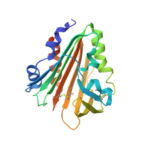
Identification and architecture of a putative secretion tube across mycobacterial outer envelope.

Cai, X.Liu, L.Qiu, C.Wen, C.He, Y.Cui, Y.Li, S.Zhang, X.Zhang, L.Tian, C.Bi, L.Zhou, Z.H.Gong, W.

(2021) Sci Adv 7

  • DOI: https://doi.org/10.1126/sciadv.abg5656
  • Primary Citation of Related Structures:  
    7CU8, 7CU9

  • PubMed Abstract: 

    Tuberculosis-causing mycobacteria have thick cell-wall and capsule layers that are formed from complex structures. Protein secretion across these barriers depends on a specialized protein secretion system, but none has been reported. We show that Mycobacterium tuberculosis Rv3705c and its homologous MSMEG_6251 in Mycobacterium smegmatis are tube-forming proteins in the mycobacterial envelope (TiME). Crystallographic and cryo-EM structures of these two proteins show that both proteins form rotationally symmetric rings. Two layers of TiME rings pack together in a tail-to-tail manner into a ring-shaped complex, which, in turn, stacks together to form tubes. M. smegmatis TiME was detected mainly in the cell wall and capsule. Knocking out the TiME gene markedly decreased the amount of secreted protein in the M. smegmatis culture medium, and expression of this gene in knocked-out strain partially restored the level of secreted protein. Our structure and functional data thus suggest that TiME forms a protein transport tube across the mycobacterial outer envelope.


  • Organizational Affiliation

    School of Life Sciences, University of Science and Technology of China, Hefei, Anhui, China.


Macromolecules
Find similar proteins by:  (by identity cutoff)  |  3D Structure
Entity ID: 1
MoleculeChains Sequence LengthOrganismDetailsImage
Tube-forming protein in Mycobacterial Envelope (TiME)
A, B, C, D, E
A, B, C, D, E, F, G
202Mycobacterium tuberculosis H37RvMutation(s): 0 
Gene Names: Rv3705c
UniProt
Find proteins for I6XI06 (Mycobacterium tuberculosis (strain ATCC 25618 / H37Rv))
Explore I6XI06 
Go to UniProtKB:  I6XI06
Entity Groups  
Sequence Clusters30% Identity50% Identity70% Identity90% Identity95% Identity100% Identity
UniProt GroupI6XI06
Sequence Annotations
Expand
  • Reference Sequence
Small Molecules
Ligands 1 Unique
IDChains Name / Formula / InChI Key2D Diagram3D Interactions
SO4
Query on SO4

Download Ideal Coordinates CCD File 
H [auth A]
I [auth A]
J [auth B]
K [auth C]
L [auth C]
H [auth A],
I [auth A],
J [auth B],
K [auth C],
L [auth C],
M [auth D],
N [auth E],
O [auth E],
P [auth F],
Q [auth G],
R [auth G]
SULFATE ION
O4 S
QAOWNCQODCNURD-UHFFFAOYSA-L
Experimental Data & Validation

Experimental Data

  • Method: X-RAY DIFFRACTION
  • Resolution: 3.30 Å
  • R-Value Free: 0.235 
  • R-Value Work: 0.223 
  • R-Value Observed: 0.223 
  • Space Group: P 61 2 2
Unit Cell:
Length ( Å )Angle ( ˚ )
a = 198.002α = 90
b = 198.002β = 90
c = 364.108γ = 120
Software Package:
Software NamePurpose
REFMACrefinement
PDB_EXTRACTdata extraction
HKL-2000data reduction
HKL-2000data scaling
PHASERphasing

Structure Validation

View Full Validation Report


Entry History & Funding Information

Deposition Data


Funding OrganizationLocationGrant Number
National Natural Science Foundation of China (NSFC)China31570841

Revision History  (Full details and data files)

  • Version 1.0: 2021-08-25
    Type: Initial release
  • Version 1.1: 2022-03-16
    Changes: Database references
  • Version 1.2: 2023-11-29
    Changes: Data collection, Refinement description