


Structure of the CYP102A1 Haem Domain with N-Carboxybenzyl-L-Prolyl-L-Phenylalanine in complex with Cyclohexylamine at 1.49 Angstrom Resolution
Shoji, O., Stanfield, J.K.To be published.
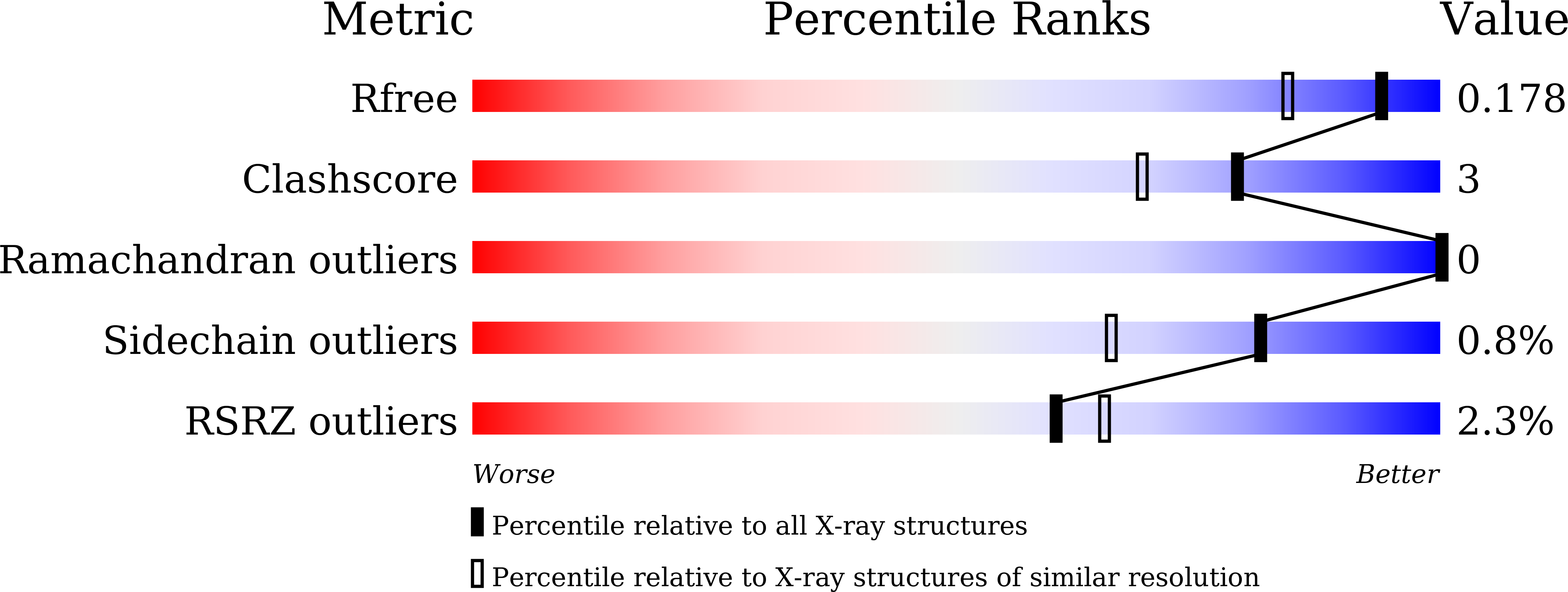
Experimental Data Snapshot
Entity ID: 1 | |||||
|---|---|---|---|---|---|
| Molecule | Chains | Sequence Length | Organism | Details | Image |
| Bifunctional cytochrome P450/NADPH--P450 reductase | 456 | Priestia megaterium | Mutation(s): 0 Gene Names: cyp102A1 EC: 1.14.14.1 (PDB Primary Data), 1.6.2.4 (PDB Primary Data) | 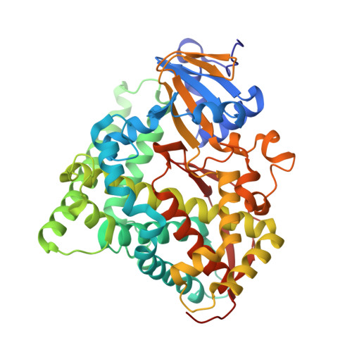 | |
UniProt | |||||
Find proteins for P14779 (Priestia megaterium (strain ATCC 14581 / DSM 32 / CCUG 1817 / JCM 2506 / NBRC 15308 / NCIMB 9376 / NCTC 10342 / NRRL B-14308 / VKM B-512 / Ford 19)) Explore P14779 Go to UniProtKB: P14779 | |||||
Entity Groups | |||||
| Sequence Clusters | 30% Identity50% Identity70% Identity90% Identity95% Identity100% Identity | ||||
| UniProt Group | P14779 | ||||
Sequence AnnotationsExpand | |||||
| |||||
| Ligands 4 Unique | |||||
|---|---|---|---|---|---|
| ID | Chains | Name / Formula / InChI Key | 2D Diagram | 3D Interactions | |
| HEM Query on HEM | C [auth A], K [auth B] | PROTOPORPHYRIN IX CONTAINING FE C34 H32 Fe N4 O4 KABFMIBPWCXCRK-RGGAHWMASA-L |  | ||
| ZP6 (Subject of Investigation/LOI) Query on ZP6 | E [auth A], M [auth B] | (2S)-3-phenyl-2-[[(2S)-1-phenylmethoxycarbonylpyrrolidin-2-yl]carbonylamino]propanoic acid C22 H24 N2 O5 GMUYSWQSSRROPG-OALUTQOASA-N |  | ||
| HAI Query on HAI | D [auth A], L [auth B] | CYCLOHEXYLAMMONIUM ION C6 H14 N PAFZNILMFXTMIY-UHFFFAOYSA-O |  | ||
| GOL Query on GOL | F [auth A] G [auth A] H [auth A] I [auth A] J [auth A] | GLYCEROL C3 H8 O3 PEDCQBHIVMGVHV-UHFFFAOYSA-N |  | ||
| Length ( Å ) | Angle ( ˚ ) |
|---|---|
| a = 58.82 | α = 90 |
| b = 128.83 | β = 90 |
| c = 149.33 | γ = 90 |
| Software Name | Purpose |
|---|---|
| REFMAC | refinement |
| XDS | data reduction |
| XSCALE | data scaling |
| MOLREP | phasing |
| PDB_EXTRACT | data extraction |
| Funding Organization | Location | Grant Number |
|---|---|---|
| Japan Science and Technology | Japan | JPMJCR15P3 |
RCSB PDB (citation) is hosted by
RCSB PDB is a member of the

