Structural and Biochemical Characterization of EFhd1/Swiprosin-2, an Actin-Binding Protein in Mitochondria.
Mun, S.A., Park, J., Park, K.R., Lee, Y., Kang, J.Y., Park, T., Jin, M., Yang, J., Jun, C.D., Eom, S.H.(2020) Front Cell Dev Biol 8: 628222-628222
- PubMed: 33537316
- DOI: https://doi.org/10.3389/fcell.2020.628222
- Primary Citation of Related Structures:
7CLT - PubMed Abstract:
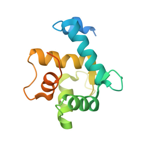
Ca 2+ regulates several cellular functions, including signaling events, energy production, and cell survival. These cellular processes are mediated by Ca 2+ -binding proteins, such as EF-hand superfamily proteins. Among the EF-hand superfamily proteins, allograft inflammatory factor-1 (AIF-1) and swiprosin-1/EF-hand domain-containing protein 2 (EFhd2) are cytosolic actin-binding proteins. AIF-1 modulates the cytoskeleton and increases the migration of immune cells. EFhd2 is also a cytoskeletal protein implicated in immune cell activation and brain cell functions. EFhd1, a mitochondrial fraternal twin of EFhd2, mediates neuronal and pro-/pre-B cell differentiation and mitoflash activation. Although EFhd1 is important for maintaining mitochondrial morphology and energy synthesis, its mechanism of action remains unclear. Here, we report the crystal structure of the EFhd1 core domain comprising a C-terminus of a proline-rich region, two EF-hand domains, and a ligand mimic helix. Structural comparisons of EFhd1, EFhd2, and AIF-1 revealed similarities in their overall structures. In the structure of the EFhd1 core domain, two Zn 2+ ions were observed at the interface of the crystal contact, suggesting the possibility of Zn 2+ -mediated multimerization. In addition, we found that EFhd1 has Ca 2+ -independent β-actin-binding and Ca 2+ -dependent β-actin-bundling activities. These findings suggest that EFhd1, an actin-binding and -bundling protein in the mitochondria, may contribute to the Ca 2+ -dependent regulation of mitochondrial morphology and energy synthesis.
Organizational Affiliation:
School of Life Sciences, Gwangju Institute of Science and Technology, Gwangju, South Korea.