7CCD

Sulfur binding domain of SprMcrA complexed with phosphorothioated DNA


Experimental Data Snapshot

  • Method: X-RAY DIFFRACTION
  • Resolution: 2.42 Å
  • R-Value Free: 0.249 
  • R-Value Work: 0.201 
  • R-Value Observed: 0.204 

wwPDB Validation   3D Report Full Report


This is version 1.3 of the entry. See complete history


Literature

DNA backbone interactions impact the sequence specificity of DNA sulfur-binding domains: revelations from structural analyses.

Yu, H.Li, J.Liu, G.Zhao, G.Wang, Y.Hu, W.Deng, Z.Wu, G.Gan, J.Zhao, Y.L.He, X.

(2020) Nucleic Acids Res 48: 8755-8766

  • DOI: https://doi.org/10.1093/nar/gkaa574
  • Primary Citation of Related Structures:  
    7CC9, 7CCD, 7CCJ

  • PubMed Abstract: 

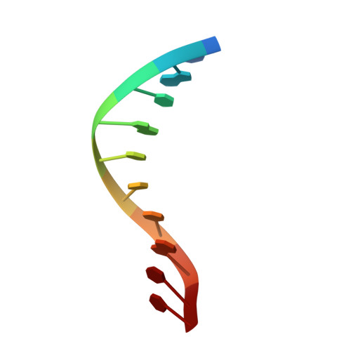
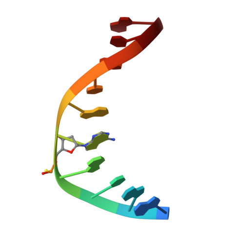
    The sulfur atom of phosphorothioated DNA (PT-DNA) is coordinated by a surface cavity in the conserved sulfur-binding domain (SBD) of type IV restriction enzymes. However, some SBDs cannot recognize the sulfur atom in some sequence contexts. To illustrate the structural determinants for sequence specificity, we resolved the structure of SBDSpr, from endonuclease SprMcrA, in complex with DNA of GPSGCC, GPSATC and GPSAAC contexts. Structural and computational analyses explained why it binds the above PT-DNAs with an affinity in a decreasing order. The structural analysis of SBDSpr-GPSGCC and SBDSco-GPSGCC, the latter only recognizes DNA of GPSGCC, revealed that a positively charged loop above the sulfur-coordination cavity electrostatically interacts with the neighboring DNA phosphate linkage. The structural analysis indicated that the DNA-protein hydrogen bonding pattern and weak non-bonded interaction played important roles in sequence specificity of SBD protein. Exchanges of the positively-charged amino acid residues with the negatively-charged residues in the loop would enable SBDSco to extend recognization for more PT-DNA sequences, implying that type IV endonucleases can be engineered to recognize PT-DNA in novel target sequences.


  • Organizational Affiliation

    State Key Laboratory of Microbial Metabolism, Joint International Research Laboratory of Metabolic & Developmental Sciences, School of Life Sciences & Biotechnology, Shanghai Jiao Tong University, 800 Dongchuan Road, Shanghai 200240, People's Republic of China.


Macromolecules

Find similar proteins by:  (by identity cutoff)  |  3D Structure
Entity ID: 1
MoleculeChains Sequence LengthOrganismDetailsImage
HNHc domain-containing proteinA,
D [auth B]
169Streptomyces pristinaespiralisMutation(s): 0 
Gene Names: SPRI_5136
UniProt
Find proteins for A0A0M4DML1 (Streptomyces pristinaespiralis)
Explore A0A0M4DML1 
Go to UniProtKB:  A0A0M4DML1
Entity Groups  
Sequence Clusters30% Identity50% Identity70% Identity90% Identity95% Identity100% Identity
UniProt GroupA0A0M4DML1
Sequence Annotations
Expand
  • Reference Sequence

Find similar nucleic acids by:  Sequence   |   3D Structure  

Entity ID: 2
MoleculeChains LengthOrganismImage
DNA (5'-D(*CP*AP*CP*GP*TP*TP*CP*GP*CP*C)-3')B [auth E],
E [auth C]
10Escherichia coli B7A
Sequence Annotations
Expand
  • Reference Sequence

Find similar nucleic acids by:  Sequence   |   3D Structure  

Entity ID: 3
MoleculeChains LengthOrganismImage
DNA (5'-D(*GP*GP*CP*GP*AS*AP*CP*GP*TP*G)-3')C [auth F],
F [auth D]
10Escherichia coli B7A
Sequence Annotations
Expand
  • Reference Sequence
Experimental Data & Validation

Experimental Data

  • Method: X-RAY DIFFRACTION
  • Resolution: 2.42 Å
  • R-Value Free: 0.249 
  • R-Value Work: 0.201 
  • R-Value Observed: 0.204 
  • Space Group: P 1
Unit Cell:
Length ( Å )Angle ( ˚ )
a = 46.94α = 107.04
b = 48.94β = 109.79
c = 56.19γ = 97.26
Software Package:
Software NamePurpose
PHENIXrefinement
SCALAdata scaling
PDB_EXTRACTdata extraction
HKL-3000data reduction
PHENIXphasing

Structure Validation

View Full Validation Report


Entry History & Funding Information

Deposition Data


Funding OrganizationLocationGrant Number
National Natural Science Foundation of China (NSFC)China31670034
National Natural Science Foundation of China (NSFC)China31470195
National Natural Science Foundation of China (NSFC)China21661140002

Revision History  (Full details and data files)

  • Version 1.0: 2020-07-08
    Type: Initial release
  • Version 1.1: 2020-08-05
    Changes: Database references
  • Version 1.2: 2021-01-20
    Changes: Database references
  • Version 1.3: 2023-11-29
    Changes: Data collection, Database references, Refinement description