7CAO

Crystal structure of red chromoprotein from Olindias formosa


Experimental Data Snapshot

  • Method: X-RAY DIFFRACTION
  • Resolution: 2.05 Å
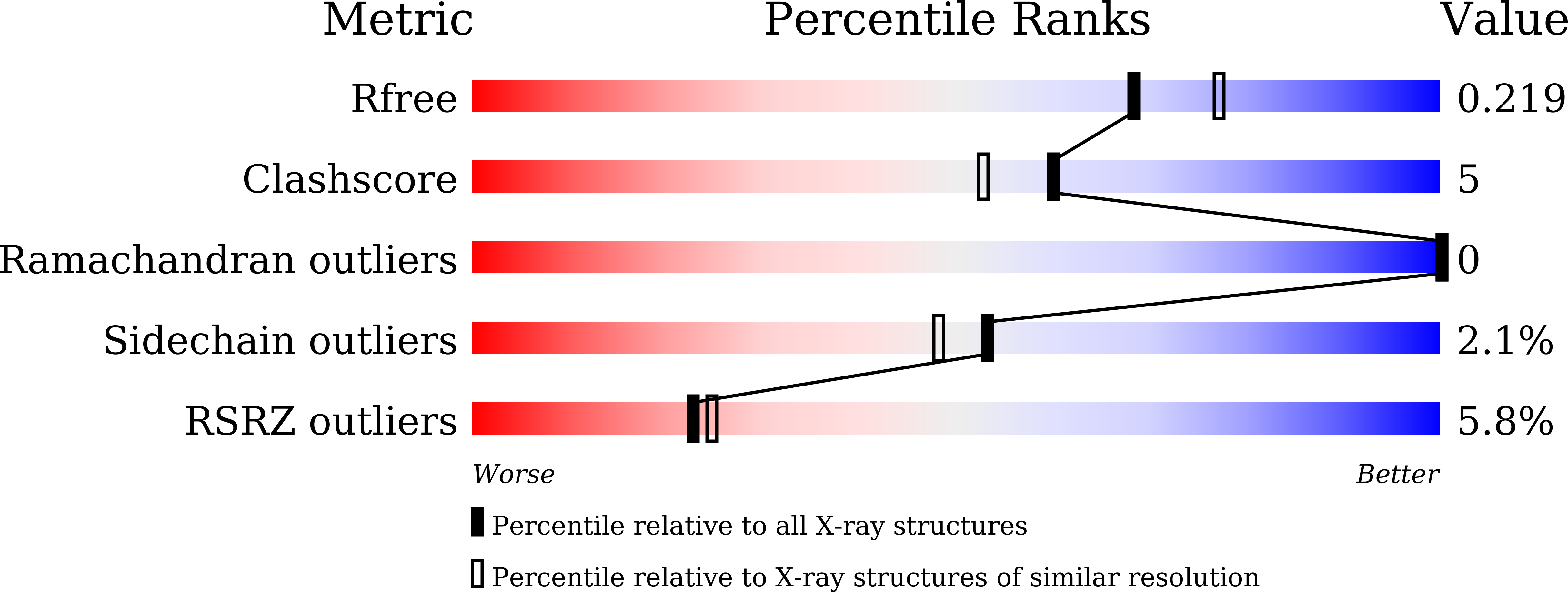
  • R-Value Free: 0.211 
  • R-Value Work: 0.192 
  • R-Value Observed: 0.193 

wwPDB Validation   3D Report Full Report


This is version 2.1 of the entry. See complete history


Literature

Structure-based analysis and evolution of a monomerized red-colored chromoprotein from the Olindias formosa jellyfish.

Zhai, L.Nakashima, R.Shinoda, H.Ike, Y.Matsuda, T.Nagai, T.

(2022) Protein Sci 31: e4285-e4285

  • DOI: https://doi.org/10.1002/pro.4285
  • Primary Citation of Related Structures:  
    7CAO

  • PubMed Abstract: 

    GFP-like chromoproteins (CPs) with non-fluorescence ability have been used as bioimaging probes. Existing CPs have voids in the optical absorption window which limits their extensibility. The development of new CP color is therefore ongoing. Here, we cloned CPs from the jellyfish, Olindias formosa, and developed a completely non-fluorescent monomeric red CP, R-Velour, with an absorption peak at 528 nm. To analyze the photophysical properties from a structural aspect, we determined the crystal structure of R-Velour at a 2.1 Å resolution. R-Velour has a trans-chromophore similar to the green fluorescence protein, Gamillus, derived from the same jellyfish. However, in contrast to the two coplanar chromophoric rings in Gamillus, R-Velour has a large torsion inducing non-fluorescence property. Through site-directed mutagenesis, we surveyed residues surrounding the chromophore and found a key residue, Ser155, which contributes to the generation of four-color variants with the bathochromic and hypsochromic shift of the absorption peak, ranging from 506 to 554 nm. The recently proposed spectrum shift theory, based on the Marcus-Hush model, supports the spectrum shift of these mutants. These findings may support further development of R-Velour variants with useful absorption characteristics for bioimaging, including fluorescence lifetime imaging and photoacoustic imaging.


  • Organizational Affiliation

    Graduate School of Frontier Bioscience, Osaka University, Suita, Japan.


Macromolecules
Find similar proteins by:  (by identity cutoff)  |  3D Structure
Entity ID: 1
MoleculeChains Sequence LengthOrganismDetailsImage
Chromoprotein262Olindias formosusMutation(s): 0 
Entity Groups  
Sequence Clusters30% Identity50% Identity70% Identity90% Identity95% Identity100% Identity
Sequence Annotations
Expand
  • Reference Sequence
Small Molecules
Modified Residues  1 Unique
IDChains TypeFormula2D DiagramParent
QYG
Query on QYG
A
L-PEPTIDE LINKINGC16 H18 N4 O5GLN, TYR, GLY
Experimental Data & Validation

Experimental Data

  • Method: X-RAY DIFFRACTION
  • Resolution: 2.05 Å
  • R-Value Free: 0.211 
  • R-Value Work: 0.192 
  • R-Value Observed: 0.193 
  • Space Group: I 41 2 2
Unit Cell:
Length ( Å )Angle ( ˚ )
a = 116.476α = 90
b = 116.476β = 90
c = 161.444γ = 90
Software Package:
Software NamePurpose
REFMACrefinement
XDSdata reduction
XSCALEdata scaling
PDB_EXTRACTdata extraction
MOLREPphasing

Structure Validation

View Full Validation Report


Entry History & Funding Information

Deposition Data


Funding OrganizationLocationGrant Number
Ministry of Education, Culture, Sports, Science and Technology (Japan)Japan18H05410

Revision History  (Full details and data files)

  • Version 1.0: 2022-04-20
    Type: Initial release
  • Version 1.1: 2022-05-11
    Changes: Database references
  • Version 1.2: 2022-05-18
    Changes: Database references
  • Version 2.0: 2023-11-15
    Changes: Atomic model, Data collection, Derived calculations
  • Version 2.1: 2023-11-29
    Changes: Refinement description