7BTK

E.coli beta-galactosidase (E537Q) in complex with fluorescent probe KSA01


Experimental Data Snapshot

  • Method: X-RAY DIFFRACTION
  • Resolution: 2.70 Å
  • R-Value Free: 0.233 
  • R-Value Work: 0.218 
  • R-Value Observed: 0.218 

wwPDB Validation   3D Report Full Report


Ligand Structure Quality Assessment 


This is version 1.3 of the entry. See complete history


Literature

Two-Dimensional Design Strategy to Construct Smart Fluorescent Probes for the Precise Tracking of Senescence.

Gao, Y.Hu, Y.Liu, Q.Li, X.Li, X.Kim, C.Y.James, T.D.Li, J.Chen, X.Guo, Y.

(2021) Angew Chem Int Ed Engl 60: 10756-10765

  • DOI: https://doi.org/10.1002/anie.202101278
  • Primary Citation of Related Structures:  
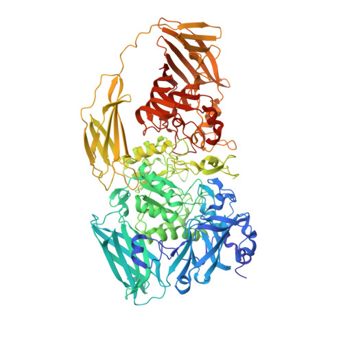
    7BRS, 7BTK

  • PubMed Abstract: 

    The tracking of cellular senescence usually depends on the detection of senescence-associated β-galactosidase (SA-β-gal). Previous probes for SA-β-gal with this purpose only cover a single dimension: the accumulation of this enzyme in lysosomes. However, this is insufficient to determine the destiny of senescence because endogenous β-gal enriched in lysosomes is not only related to senescence, but also to some other physiological processes. To address this issue, we introduce our fluorescent probes including a second dimension: lysosomal pH, since de-acidification is a unique feature of the lysosomes in senescent cells. With this novel design, our probes achieved excellent discrimination of SA-β-gal from cancer-associated β-gal, which enables them to track cellular senescence as well as tissue aging more precisely. Our crystal structures of a model enzyme E. coli β-gal mutant (E537Q) complexed with each probe further revealed the structural basis for probe recognition.


  • Organizational Affiliation

    Key Laboratory of Synthetic and Natural Functional Molecule of the Ministry of Education, College of Chemistry and Materials Science, Northwest University, Xi'an, 710127, China.


Macromolecules
Find similar proteins by:  (by identity cutoff)  |  3D Structure
Entity ID: 1
MoleculeChains Sequence LengthOrganismDetailsImage
Beta-galactosidase
A, B, C, D
1,025Escherichia coli K-12Mutation(s): 1 
Gene Names: lacZ
EC: 3.2.1.23
UniProt
Find proteins for P00722 (Escherichia coli (strain K12))
Explore P00722 
Go to UniProtKB:  P00722
Entity Groups  
Sequence Clusters30% Identity50% Identity70% Identity90% Identity95% Identity100% Identity
UniProt GroupP00722
Sequence Annotations
Expand
  • Reference Sequence
Small Molecules
Ligands 5 Unique
IDChains Name / Formula / InChI Key2D Diagram3D Interactions
F6L (Subject of Investigation/LOI)
Query on F6L

Download Ideal Coordinates CCD File 
E [auth A],
JA [auth C],
V [auth B],
WA [auth D]
4-[[2-[(E)-2-[4-[(2S,3R,4S,5R,6R)-6-(hydroxymethyl)-3,4,5-tris(oxidanyl)oxan-2-yl]oxyphenyl]ethenyl]-3,3-dimethyl-2H-indol-1-yl]methyl]benzoic acid
C32 H34 N O8
ZRXNUVMWWIBYNP-BARJCIPKSA-O
GOL
Query on GOL

Download Ideal Coordinates CCD File 
IA [auth B]GLYCEROL
C3 H8 O3
PEDCQBHIVMGVHV-UHFFFAOYSA-N
DMS
Query on DMS

Download Ideal Coordinates CCD File 
AA [auth B]
BA [auth B]
BB [auth D]
CA [auth B]
CB [auth D]
AA [auth B],
BA [auth B],
BB [auth D],
CA [auth B],
CB [auth D],
DA [auth B],
DB [auth D],
EA [auth B],
EB [auth D],
FA [auth B],
FB [auth D],
GA [auth B],
GB [auth D],
HA [auth B],
HB [auth D],
IB [auth D],
J [auth A],
K [auth A],
L [auth A],
M [auth A],
N [auth A],
O [auth A],
OA [auth C],
P [auth A],
PA [auth C],
Q [auth A],
QA [auth C],
R [auth A],
RA [auth C],
S [auth A],
SA [auth C],
T [auth A],
TA [auth C],
U [auth A],
UA [auth C],
VA [auth C],
Y [auth B],
Z [auth B]
DIMETHYL SULFOXIDE
C2 H6 O S
IAZDPXIOMUYVGZ-UHFFFAOYSA-N
MG
Query on MG

Download Ideal Coordinates CCD File 
F [auth A]
G [auth A]
KA [auth C]
LA [auth C]
W [auth B]
F [auth A],
G [auth A],
KA [auth C],
LA [auth C],
W [auth B],
XA [auth D],
YA [auth D]
MAGNESIUM ION
Mg
JLVVSXFLKOJNIY-UHFFFAOYSA-N
NA
Query on NA

Download Ideal Coordinates CCD File 
AB [auth D]
H [auth A]
I [auth A]
MA [auth C]
NA [auth C]
AB [auth D],
H [auth A],
I [auth A],
MA [auth C],
NA [auth C],
X [auth B],
ZA [auth D]
SODIUM ION
Na
FKNQFGJONOIPTF-UHFFFAOYSA-N
Experimental Data & Validation

Experimental Data

  • Method: X-RAY DIFFRACTION
  • Resolution: 2.70 Å
  • R-Value Free: 0.233 
  • R-Value Work: 0.218 
  • R-Value Observed: 0.218 
  • Space Group: C 1 2 1
Unit Cell:
Length ( Å )Angle ( ˚ )
a = 249.832α = 90
b = 85.454β = 94.15
c = 243.307γ = 90
Software Package:
Software NamePurpose
REFMACrefinement
PDB_EXTRACTdata extraction
XDSdata reduction
Aimlessdata scaling
MOLREPphasing

Structure Validation

View Full Validation Report


Ligand Structure Quality Assessment 


Entry History & Funding Information

Deposition Data


Funding OrganizationLocationGrant Number
National Natural Science Foundation of China (NSFC)China21977082
National Natural Science Foundation of China (NSFC)China21472148
National Natural Science Foundation of China (NSFC)China21807088
National Natural Science Foundation of China (NSFC)China81872747

Revision History  (Full details and data files)

  • Version 1.0: 2021-03-03
    Type: Initial release
  • Version 1.1: 2021-03-10
    Changes: Database references
  • Version 1.2: 2021-05-12
    Changes: Database references
  • Version 1.3: 2023-11-29
    Changes: Data collection, Database references, Refinement description