7BRD

Crystal structure of Peptidyl-tRNA hydrolase from Klebsiella pneumoniae


Experimental Data Snapshot

  • Method: X-RAY DIFFRACTION
  • Resolution: 1.89 Å
  • R-Value Free: 0.222 
  • R-Value Work: 0.174 

wwPDB Validation   3D Report Full Report


This is version 1.1 of the entry. See complete history


Literature

Structural and functional characterization of peptidyl-tRNA hydrolase from Klebsiella pneumoniae.

Mundra, S.Pal, R.K.Tripathi, S.Jain, A.Arora, A.

(2021) Biochim Biophys Acta Proteins Proteom 1869: 140554-140554

  • DOI: https://doi.org/10.1016/j.bbapap.2020.140554
  • Primary Citation of Related Structures:  
    7BRD

  • PubMed Abstract: 

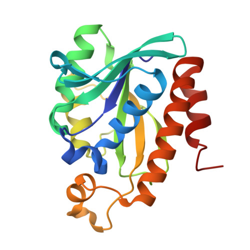
    Klebsiella pneumoniae is a member of the ESKAPE panel of pathogens that are top priority to tackle AMR. Bacterial peptidyl tRNA hydrolase (Pth), an essential, ubiquitous enzyme, hydrolyzes the peptidyl-tRNAs that accumulate in the cytoplasm because of premature termination of translation. Pth cleaves the ester bond between 2' or 3' hydroxyl of the ribose in the tRNA and C-terminal carboxylate of the peptide, thereby making free tRNA available for repeated cycles of protein synthesis and preventing cell death by alleviating tRNA starvation. Pth structures have been determined in peptide-bound or peptide-free states. In peptide-bound state, highly conserved residues F67, N69 and N115 adopt a conformation that is conducive to their interaction with peptide moiety of the substrate. While, in peptide-free state, these residues move away from the catalytic center, perhaps, in order to facilitate release of hydrolysed peptide. Here, we present a novel X-ray crystal structure of Pth from Klebsiella pneumoniae (KpPth), at 1.89 Å resolution, in which out of the two molecules in the asymmetric unit, one reflects the peptide-bound while the other reflects peptide-free conformation of the conserved catalytic site residues. Each molecule of the protein has canonical structure with seven stranded β-sheet structure surrounded by six α-helices. MD simulations indicate that both the forms converge over 500 ns simulation to structures with wider opening of the crevice at peptide-binding end. In solution, KpPth is monomeric and its 2D-HSQC spectrum displays a single set of well dispersed peaks. Further, KpPth was demonstrated to be enzymatically active on BODIPY-Lys-tRNA Lys3 .


  • Organizational Affiliation

    Molecular and Structural Biology Division, CSIR-Central Drug Research Institute, Lucknow 226031, India; Department of Science and Technology, New Delhi 110016, India.


Macromolecules
Find similar proteins by:  (by identity cutoff)  |  3D Structure
Entity ID: 1
MoleculeChains Sequence LengthOrganismDetailsImage
Peptidyl-tRNA hydrolase
A, B
194Klebsiella pneumoniaeMutation(s): 0 
Gene Names: pthBL124_00016980EAO17_03025F3P37_02700SAMEA3538476_04832SK89_03098
EC: 3.1.1.29
UniProt
Find proteins for A6TAP7 (Klebsiella pneumoniae subsp. pneumoniae (strain ATCC 700721 / MGH 78578))
Explore A6TAP7 
Go to UniProtKB:  A6TAP7
Entity Groups  
Sequence Clusters30% Identity50% Identity70% Identity90% Identity95% Identity100% Identity
UniProt GroupA6TAP7
Sequence Annotations
Expand
  • Reference Sequence
Small Molecules
Ligands 7 Unique
IDChains Name / Formula / InChI Key2D Diagram3D Interactions
PGE
Query on PGE

Download Ideal Coordinates CCD File 
C [auth A]TRIETHYLENE GLYCOL
C6 H14 O4
ZIBGPFATKBEMQZ-UHFFFAOYSA-N
PEG
Query on PEG

Download Ideal Coordinates CCD File 
D [auth A],
V [auth B]
DI(HYDROXYETHYL)ETHER
C4 H10 O3
MTHSVFCYNBDYFN-UHFFFAOYSA-N
SO4
Query on SO4

Download Ideal Coordinates CCD File 
DA [auth B],
EA [auth B],
S [auth A],
T [auth A]
SULFATE ION
O4 S
QAOWNCQODCNURD-UHFFFAOYSA-L
BME
Query on BME

Download Ideal Coordinates CCD File 
Z [auth B]BETA-MERCAPTOETHANOL
C2 H6 O S
DGVVWUTYPXICAM-UHFFFAOYSA-N
EDO
Query on EDO

Download Ideal Coordinates CCD File 
AA [auth B]
E [auth A]
F [auth A]
K [auth A]
L [auth A]
AA [auth B],
E [auth A],
F [auth A],
K [auth A],
L [auth A],
W [auth B],
X [auth B],
Y [auth B]
1,2-ETHANEDIOL
C2 H6 O2
LYCAIKOWRPUZTN-UHFFFAOYSA-N
ACT
Query on ACT

Download Ideal Coordinates CCD File 
BA [auth B]
CA [auth B]
G [auth A]
H [auth A]
I [auth A]
BA [auth B],
CA [auth B],
G [auth A],
H [auth A],
I [auth A],
J [auth A],
M [auth A],
N [auth A],
U [auth A]
ACETATE ION
C2 H3 O2
QTBSBXVTEAMEQO-UHFFFAOYSA-M
CL
Query on CL

Download Ideal Coordinates CCD File 
O [auth A],
P [auth A],
Q [auth A],
R [auth A]
CHLORIDE ION
Cl
VEXZGXHMUGYJMC-UHFFFAOYSA-M
Experimental Data & Validation

Experimental Data

  • Method: X-RAY DIFFRACTION
  • Resolution: 1.89 Å
  • R-Value Free: 0.222 
  • R-Value Work: 0.174 
  • Space Group: P 21 21 21
Unit Cell:
Length ( Å )Angle ( ˚ )
a = 70.203α = 90
b = 72.372β = 90
c = 77.415γ = 90
Software Package:
Software NamePurpose
REFMACrefinement
HKL-2000data reduction
HKL-2000data scaling
PHASERphasing

Structure Validation

View Full Validation Report


Entry History & Funding Information

Deposition Data


Funding OrganizationLocationGrant Number
Department of Biotechnology (DBT, India)IndiaGAP328

Revision History  (Full details and data files)

  • Version 1.0: 2021-03-17
    Type: Initial release
  • Version 1.1: 2023-11-29
    Changes: Data collection, Database references, Derived calculations, Refinement description