7BE4

Crystal structure of MAP kinase p38 alpha in complex with inhibitor SR159


Experimental Data Snapshot

  • Method: X-RAY DIFFRACTION
  • Resolution: 2.10 Å
  • R-Value Free: 0.268 
  • R-Value Work: 0.215 
  • R-Value Observed: 0.217 

wwPDB Validation   3D Report Full Report


Ligand Structure Quality Assessment 


This is version 1.2 of the entry. See complete history


Literature

Development of a Selective Dual Discoidin Domain Receptor (DDR)/p38 Kinase Chemical Probe.

Rohm, S.Berger, B.T.Schroder, M.Chatterjee, D.Mathea, S.Joerger, A.C.Pinkas, D.M.Bufton, J.C.Tjaden, A.Kovooru, L.Kudolo, M.Pohl, C.Bullock, A.N.Muller, S.Laufer, S.Knapp, S.

(2021) J Med Chem 64: 13451-13474

  • DOI: https://doi.org/10.1021/acs.jmedchem.1c00868
  • Primary Citation of Related Structures:  
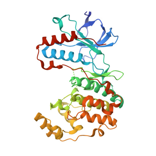
    7BCM, 7BDO, 7BDQ, 7BE4, 7BE5, 7BE6

  • PubMed Abstract: 

    Discoidin domain receptors 1 and 2 (DDR1/2) play a central role in fibrotic disorders, such as renal and pulmonary fibrosis, atherosclerosis, and various forms of cancer. Potent and selective inhibitors, so-called chemical probe compounds, have been developed to study DDR1/2 kinase signaling. However, these inhibitors showed undesired activity on other kinases such as the tyrosine protein kinase receptor TIE or tropomyosin receptor kinases, which are related to angiogenesis and neuronal toxicity. In this study, we optimized our recently published p38 mitogen-activated protein kinase inhibitor 7 toward a potent and cell-active dual DDR/p38 chemical probe and developed a structurally related negative control. The structure-guided design approach used provided insights into the P-loop folding process of p38 and how targeting of non-conserved amino acids modulates inhibitor selectivity. The developed and comprehensively characterized DDR/p38 probe, 30 (SR-302), is a valuable tool for studying the role of DDR kinase in normal physiology and in disease development.


  • Organizational Affiliation

    Institute of Pharmaceutical Chemistry, Johann Wolfgang Goethe University, Max-von-Laue-Str. 9, 60438 Frankfurt am Main, Germany.


Macromolecules
Find similar proteins by:  (by identity cutoff)  |  3D Structure
Entity ID: 1
MoleculeChains Sequence LengthOrganismDetailsImage
Mitogen-activated protein kinase 14361Mus musculusMutation(s): 0 
Gene Names: Mapk14Crk1Csbp1Csbp2
EC: 2.7.11.24
UniProt
Find proteins for P47811 (Mus musculus)
Explore P47811 
Go to UniProtKB:  P47811
Entity Groups  
Sequence Clusters30% Identity50% Identity70% Identity90% Identity95% Identity100% Identity
UniProt GroupP47811
Sequence Annotations
Expand
  • Reference Sequence
Small Molecules
Ligands 1 Unique
IDChains Name / Formula / InChI Key2D Diagram3D Interactions
TK5 (Subject of Investigation/LOI)
Query on TK5

Download Ideal Coordinates CCD File 
B [auth A]5-azanyl-~{N}-[[4-[[(2~{S})-4-cyclohexyl-1-[[(3~{R})-1-methylsulfonylpiperidin-3-yl]amino]-1-oxidanylidene-butan-2-yl]carbamoyl]phenyl]methyl]-1-phenyl-pyrazole-4-carboxamide
C34 H45 N7 O5 S
GODYJVJUBKYVOU-OFSOJUDTSA-N
Experimental Data & Validation

Experimental Data

  • Method: X-RAY DIFFRACTION
  • Resolution: 2.10 Å
  • R-Value Free: 0.268 
  • R-Value Work: 0.215 
  • R-Value Observed: 0.217 
  • Space Group: P 21 21 21
Unit Cell:
Length ( Å )Angle ( ˚ )
a = 66.46α = 90
b = 75.24β = 90
c = 78.19γ = 90
Software Package:
Software NamePurpose
PHENIXrefinement
PHASERphasing

Structure Validation

View Full Validation Report


Ligand Structure Quality Assessment 


Entry History 

Deposition Data

Revision History  (Full details and data files)

  • Version 1.0: 2021-03-03
    Type: Initial release
  • Version 1.1: 2022-09-14
    Changes: Database references
  • Version 1.2: 2024-01-31
    Changes: Data collection, Refinement description