7BD7

X-ray structure of Arsenoplatin-1-encapsulated horse spleen ferritin


Experimental Data Snapshot

  • Method: X-RAY DIFFRACTION
  • Resolution: 1.50 Å
  • R-Value Free: 0.185 
  • R-Value Work: 0.166 
  • R-Value Observed: 0.167 

wwPDB Validation   3D Report Full Report


Ligand Structure Quality Assessment 


This is version 1.1 of the entry. See complete history


Literature

Arsenoplatin-Ferritin Nanocage: Structure and Cytotoxicity.

Ferraro, G.Pratesi, A.Cirri, D.Imbimbo, P.Maria Monti, D.Messori, L.Merlino, A.

(2021) Int J Mol Sci 22

  • DOI: https://doi.org/10.3390/ijms22041874
  • Primary Citation of Related Structures:  
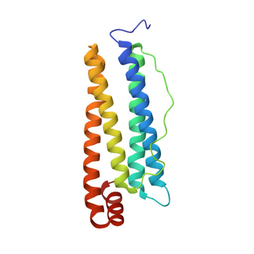
    7BD7

  • PubMed Abstract: 

    Arsenoplatin-1 (AP-1), the prototype of a novel class of metallodrugs containing a PtAs(OH) 2 core, was encapsulated within the apoferritin (AFt) nanocage. UV-Vis absorption spectroscopy and inductively coupled plasma-atomic emission spectroscopy measurements confirmed metallodrug encapsulation and allowed us to determine the average amount of AP-1 trapped inside the cage. The X-ray structure of AP-1-encapsulated AFt was solved at 1.50 Å. Diffraction data revealed that an AP-1 fragment coordinates the side chain of a His residue. The biological activity of AP-1-loaded AFt was comparatively tested on a few representative cancer and non-cancer cell lines. Even though the presence of the cage reduces the overall cytotoxicity of AP-1, it improves its selectivity towards cancer cells.


  • Organizational Affiliation

    Department of Chemistry "Ugo Schiff", University of Florence, Via Della Lastruccia, 3-13, Sesto Fiorentino, 50019 Florence, Italy.


Macromolecules
Find similar proteins by:  (by identity cutoff)  |  3D Structure
Entity ID: 1
MoleculeChains Sequence LengthOrganismDetailsImage
Ferritin light chain172Equus caballusMutation(s): 0 
UniProt
Find proteins for P02791 (Equus caballus)
Explore P02791 
Go to UniProtKB:  P02791
Entity Groups  
Sequence Clusters30% Identity50% Identity70% Identity90% Identity95% Identity100% Identity
UniProt GroupP02791
Sequence Annotations
Expand
  • Reference Sequence
Small Molecules
Ligands 6 Unique
IDChains Name / Formula / InChI Key2D Diagram3D Interactions
A6R (Subject of Investigation/LOI)
Query on A6R

Download Ideal Coordinates CCD File 
X [auth A]arsenoplatin-1
C4 H8 As N2 O4 Pt
OPINMBLEOXZNNE-UHFFFAOYSA-N
CD
Query on CD

Download Ideal Coordinates CCD File 
E [auth A]
F [auth A]
G [auth A]
H [auth A]
I [auth A]
E [auth A],
F [auth A],
G [auth A],
H [auth A],
I [auth A],
J [auth A],
K [auth A],
L [auth A],
M [auth A],
N [auth A],
O [auth A],
P [auth A],
Q [auth A],
R [auth A],
S [auth A]
CADMIUM ION
Cd
WLZRMCYVCSSEQC-UHFFFAOYSA-N
SO4
Query on SO4

Download Ideal Coordinates CCD File 
V [auth A]SULFATE ION
O4 S
QAOWNCQODCNURD-UHFFFAOYSA-L
GOL
Query on GOL

Download Ideal Coordinates CCD File 
T [auth A],
U [auth A]
GLYCEROL
C3 H8 O3
PEDCQBHIVMGVHV-UHFFFAOYSA-N
CL
Query on CL

Download Ideal Coordinates CCD File 
W [auth A]CHLORIDE ION
Cl
VEXZGXHMUGYJMC-UHFFFAOYSA-M
NA
Query on NA

Download Ideal Coordinates CCD File 
B [auth A],
C [auth A],
D [auth A]
SODIUM ION
Na
FKNQFGJONOIPTF-UHFFFAOYSA-N
Experimental Data & Validation

Experimental Data

  • Method: X-RAY DIFFRACTION
  • Resolution: 1.50 Å
  • R-Value Free: 0.185 
  • R-Value Work: 0.166 
  • R-Value Observed: 0.167 
  • Space Group: F 4 3 2
Unit Cell:
Length ( Å )Angle ( ˚ )
a = 180.91α = 90
b = 180.91β = 90
c = 180.91γ = 90
Software Package:
Software NamePurpose
REFMACrefinement
PDB_EXTRACTdata extraction
iMOSFLMdata reduction
STARANISOdata scaling
PHASERphasing

Structure Validation

View Full Validation Report


Ligand Structure Quality Assessment 


Entry History & Funding Information

Deposition Data


Funding OrganizationLocationGrant Number
Italian Association for Cancer ResearchItaly22587

Revision History  (Full details and data files)

  • Version 1.0: 2021-03-17
    Type: Initial release
  • Version 1.1: 2024-01-31
    Changes: Data collection, Database references, Refinement description