7B8I

Tetragonal structure of human protein kinase CK2 catalytic subunit in complex with a heparin oligo saccharide


Experimental Data Snapshot

  • Method: X-RAY DIFFRACTION
  • Resolution: 2.55 Å
  • R-Value Free: 0.240 
  • R-Value Work: 0.204 
  • R-Value Observed: 0.206 

wwPDB Validation   3D Report Full Report


This is version 1.3 of the entry. See complete history


Literature

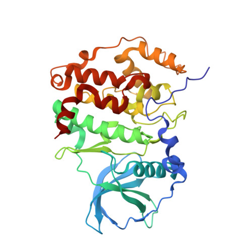
Structural basis for the design of bisubstrate inhibitors of protein kinase CK2 provided by complex structures with the substrate-competitive inhibitor heparin.

Schnitzler, A.Niefind, K.

(2021) Eur J Med Chem 214: 113223-113223

  • DOI: https://doi.org/10.1016/j.ejmech.2021.113223
  • Primary Citation of Related Structures:  
    7B8H, 7B8I

  • PubMed Abstract: 

    The Ser/Thr kinase CK2, a member of the superfamily of eukaryotic protein kinases, has an acidophilic substrate profile with the substrate recognition sequence S/T-D/E-X-D/E, and it is inhibited by polyanionic substances like heparin. The latter, a highly sulphated glucosamino glycan composed mainly of repeating 2-O-sulpho-α-l-idopyranuronic acid/N,O6-disulpho-α-d-glucosamine disaccharide units, is the longest known substrate-competitive CK2 inhibitor. The structural basis of CK2's preference for anionic substrates and substrate-competitive inhibitors is only vaguely known which limits the value of the substrate-binding region for the structure-based development of CK2 bisubstrate inhibitors. Here, a tetragonal and a monoclinic co-crystal structure of CK2α, the catalytic subunit of CK2, with a decameric heparin fragment are described. In the tetragonal structure, the heparin molecule binds to the polybasic stretch at the beginning of CK2α's helix αC, whereas in the monoclinic structure it occupies the central substrate-recognition region around the P+1 loop. Together, the structures rationalize the inhibitory efficacy of heparin fragments as a function of chain length. The monoclinic CK2α/heparin structure, in which the heparin fragment is particularly well defined, is the first CK2 structure with an anionic inhibitor of considerable size at the central part of the substrate-recognition site. The bound heparin fragment is so close to the binding site of ATP-competitive inhibitors that it can guide the design of linkers and pave the way to efficient CK2 bisubstrate inhibitors in the future.


  • Organizational Affiliation

    Universität zu Köln, Department für Chemie, Institut für Biochemie, Zülpicher Straße 47, D-50674 Köln, Germany.


Macromolecules
Find similar proteins by:  (by identity cutoff)  |  3D Structure
Entity ID: 1
MoleculeChains Sequence LengthOrganismDetailsImage
Casein kinase II subunit alpha
A, B
335Homo sapiensMutation(s): 0 
Gene Names: CSNK2A1CK2A1
EC: 2.7.11.1
UniProt & NIH Common Fund Data Resources
Find proteins for P68400 (Homo sapiens)
Explore P68400 
Go to UniProtKB:  P68400
PHAROS:  P68400
GTEx:  ENSG00000101266 
Entity Groups  
Sequence Clusters30% Identity50% Identity70% Identity90% Identity95% Identity100% Identity
UniProt GroupP68400
Sequence Annotations
Expand
  • Reference Sequence
Oligosaccharides

Help

Entity ID: 2
MoleculeChains Length2D Diagram Glycosylation3D Interactions
2-deoxy-6-O-sulfo-2-(sulfoamino)-alpha-D-glucopyranose-(1-4)-2-O-sulfo-alpha-L-idopyranuronic acid-(1-4)-2-deoxy-6-O-sulfo-2-(sulfoamino)-alpha-D-glucopyranose-(1-4)-2-O-sulfo-alpha-L-idopyranuronic acid-(1-4)-2-deoxy-6-O-sulfo-2-(sulfoamino)-alpha-D-glucopyranose-(1-4)-2-O-sulfo-alpha-L-idopyranuronic acid-(1-4)-2-deoxy-6-O-sulfo-2-(sulfoamino)-alpha-D-glucopyranose-(1-4)-2-O-sulfo-alpha-L-idopyranuronic acid
C
8N/A
Glycosylation Resources
GlyTouCan:  G22032RS
GlyCosmos:  G22032RS
Experimental Data & Validation

Experimental Data

  • Method: X-RAY DIFFRACTION
  • Resolution: 2.55 Å
  • R-Value Free: 0.240 
  • R-Value Work: 0.204 
  • R-Value Observed: 0.206 
  • Space Group: P 43 21 2
Unit Cell:
Length ( Å )Angle ( ˚ )
a = 128.401α = 90
b = 128.401β = 90
c = 124.154γ = 90
Software Package:
Software NamePurpose
PHENIXrefinement
XDSdata reduction
XSCALEdata scaling
PHASERphasing

Structure Validation

View Full Validation Report


Entry History & Funding Information

Deposition Data


Funding OrganizationLocationGrant Number
German Research Foundation (DFG)GermanyNI 643/4-1
German Research Foundation (DFG)GermanyNI 643/4-2

Revision History  (Full details and data files)

  • Version 1.0: 2021-02-03
    Type: Initial release
  • Version 1.1: 2021-02-17
    Changes: Database references
  • Version 1.2: 2021-02-24
    Changes: Database references
  • Version 1.3: 2024-01-31
    Changes: Data collection, Database references, Refinement description