7B7N

Human herpesvirus-8 gH/gL in complex with EphA2


Experimental Data Snapshot

  • Method: X-RAY DIFFRACTION
  • Resolution: 2.69 Å
  • R-Value Free: 0.265 
  • R-Value Work: 0.223 
  • R-Value Observed: 0.225 

wwPDB Validation   3D Report Full Report


This is version 1.2 of the entry. See complete history


Literature

Human herpesvirus 8 molecular mimicry of ephrin ligands facilitates cell entry and triggers EphA2 signaling.

Light, T.P.Brun, D.Guardado-Calvo, P.Pederzoli, R.Haouz, A.Neipel, F.Rey, F.A.Hristova, K.Backovic, M.

(2021) PLoS Biol 19: e3001392-e3001392

  • DOI: https://doi.org/10.1371/journal.pbio.3001392
  • Primary Citation of Related Structures:  
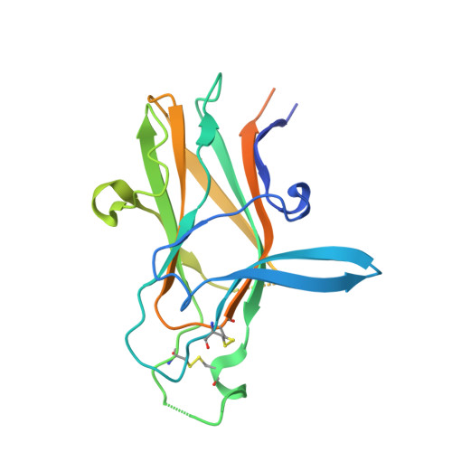
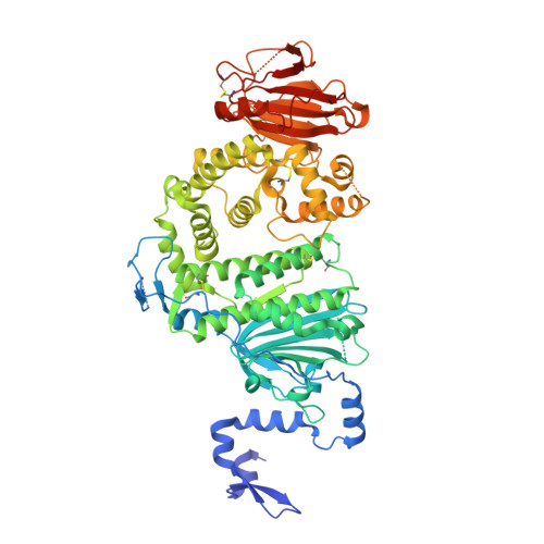
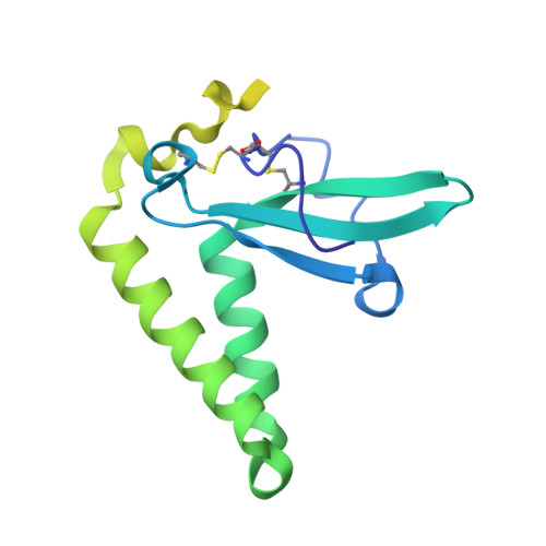
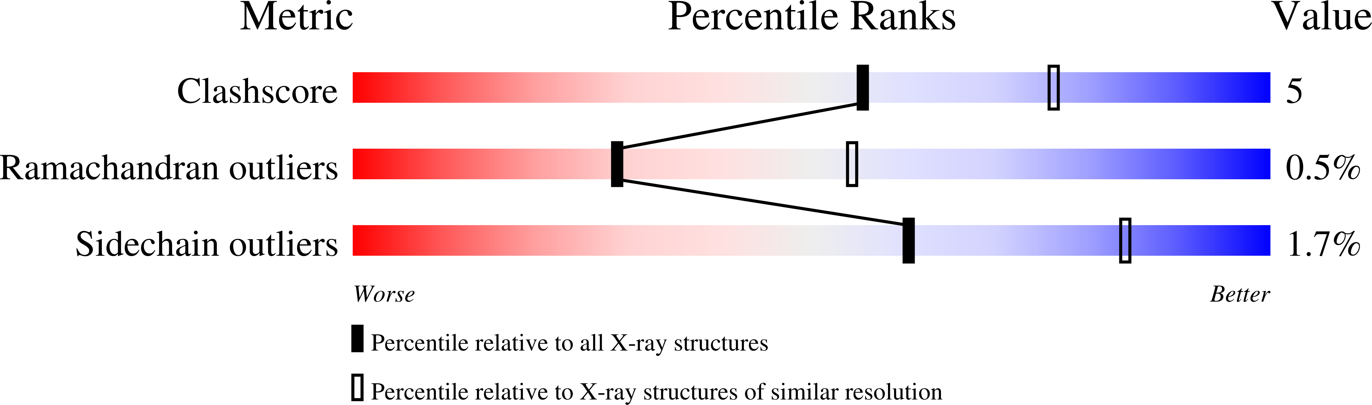
    7B7N

  • PubMed Abstract: 

    Human herpesvirus 8 (HHV-8) is an oncogenic virus that enters cells by fusion of the viral and endosomal cellular membranes in a process mediated by viral surface glycoproteins. One of the cellular receptors hijacked by HHV-8 to gain access to cells is the EphA2 tyrosine kinase receptor, and the mechanistic basis of EphA2-mediated viral entry remains unclear. Using X-ray structure analysis, targeted mutagenesis, and binding studies, we here show that the HHV-8 envelope glycoprotein complex H and L (gH/gL) binds with subnanomolar affinity to EphA2 via molecular mimicry of the receptor's cellular ligands, ephrins (Eph family receptor interacting proteins), revealing a pivotal role for the conserved gH residue E52 and the amino-terminal peptide of gL. Using FSI-FRET and cell contraction assays, we further demonstrate that the gH/gL complex also functionally mimics ephrin ligand by inducing EphA2 receptor association via its dimerization interface, thus triggering receptor signaling for cytoskeleton remodeling. These results now provide novel insight into the entry mechanism of HHV-8, opening avenues for the search of therapeutic agents that could interfere with HHV-8-related diseases.


  • Organizational Affiliation

    Department of Materials Science and Engineering, Johns Hopkins University, Baltimore, Maryland, United States of America.


Macromolecules
Find similar proteins by:  (by identity cutoff)  |  3D Structure
Entity ID: 1
MoleculeChains Sequence LengthOrganismDetailsImage
Ephrin type-A receptor 2A [auth E]215Homo sapiensMutation(s): 0 
Gene Names: EPHA2ECK
EC: 2.7.10.1
UniProt & NIH Common Fund Data Resources
Find proteins for P29317 (Homo sapiens)
Explore P29317 
Go to UniProtKB:  P29317
PHAROS:  P29317
GTEx:  ENSG00000142627 
Entity Groups  
Sequence Clusters30% Identity50% Identity70% Identity90% Identity95% Identity100% Identity
UniProt GroupP29317
Sequence Annotations
Expand
  • Reference Sequence
Find similar proteins by:  (by identity cutoff)  |  3D Structure
Entity ID: 2
MoleculeChains Sequence LengthOrganismDetailsImage
Envelope glycoprotein HB [auth H]681Human gammaherpesvirus 8Mutation(s): 0 
Gene Names: ORF22gHHHV8GK18_gp26
UniProt
Find proteins for Q98142 (Human herpesvirus 8)
Explore Q98142 
Go to UniProtKB:  Q98142
Entity Groups  
Sequence Clusters30% Identity50% Identity70% Identity90% Identity95% Identity100% Identity
UniProt GroupQ98142
Sequence Annotations
Expand
  • Reference Sequence
Find similar proteins by:  (by identity cutoff)  |  3D Structure
Entity ID: 3
MoleculeChains Sequence LengthOrganismDetailsImage
Envelope glycoprotein LC [auth L]184Human gammaherpesvirus 8Mutation(s): 1 
Gene Names: ORF47gLORF46HHV8GK18_gp49
UniProt
Find proteins for F5HDB7 (Human herpesvirus 8 type P (isolate GK18))
Explore F5HDB7 
Go to UniProtKB:  F5HDB7
Entity Groups  
Sequence Clusters30% Identity50% Identity70% Identity90% Identity95% Identity100% Identity
UniProt GroupF5HDB7
Sequence Annotations
Expand
  • Reference Sequence
Oligosaccharides

Help

Entity ID: 4
MoleculeChains Length2D Diagram Glycosylation3D Interactions
2-acetamido-2-deoxy-beta-D-glucopyranose-(1-4)-2-acetamido-2-deoxy-beta-D-glucopyranoseD [auth A],
E [auth B]
2N-Glycosylation
Glycosylation Resources
GlyTouCan:  G42666HT
GlyCosmos:  G42666HT
GlyGen:  G42666HT
Small Molecules
Ligands 3 Unique
IDChains Name / Formula / InChI Key2D Diagram3D Interactions
NAG
Query on NAG

Download Ideal Coordinates CCD File 
K [auth H],
Q [auth L]
2-acetamido-2-deoxy-beta-D-glucopyranose
C8 H15 N O6
OVRNDRQMDRJTHS-FMDGEEDCSA-N
CL
Query on CL

Download Ideal Coordinates CCD File 
F [auth E],
G [auth E],
H [auth E],
R [auth L]
CHLORIDE ION
Cl
VEXZGXHMUGYJMC-UHFFFAOYSA-M
NA
Query on NA

Download Ideal Coordinates CCD File 
I [auth E]
J [auth E]
L [auth H]
M [auth H]
N [auth H]
I [auth E],
J [auth E],
L [auth H],
M [auth H],
N [auth H],
O [auth H],
P [auth H],
S [auth L],
T [auth L],
U [auth L]
SODIUM ION
Na
FKNQFGJONOIPTF-UHFFFAOYSA-N
Experimental Data & Validation

Experimental Data

  • Method: X-RAY DIFFRACTION
  • Resolution: 2.69 Å
  • R-Value Free: 0.265 
  • R-Value Work: 0.223 
  • R-Value Observed: 0.225 
  • Space Group: C 2 2 21
Unit Cell:
Length ( Å )Angle ( ˚ )
a = 72.94α = 90
b = 129β = 90
c = 267.69γ = 90
Software Package:
Software NamePurpose
XDSdata reduction
Aimlessdata scaling
PHASERphasing
PHENIXphasing
PHENIXrefinement
BUSTERrefinement
PHENIXmodel building
Cootmodel building

Structure Validation

View Full Validation Report


Entry History & Funding Information

Deposition Data


Funding OrganizationLocationGrant Number
Pasteur InstituteFranceRecurrent funding

Revision History  (Full details and data files)

  • Version 1.0: 2020-12-30
    Type: Initial release
  • Version 1.1: 2021-09-22
    Changes: Data collection, Database references
  • Version 1.2: 2024-01-31
    Changes: Data collection, Refinement description