7B0C

[4Fe-4S]-NsrR complexed to 23-bp HmpA1 operator fragment


Experimental Data Snapshot

  • Method: X-RAY DIFFRACTION
  • Resolution: 3.00 Å
  • R-Value Free: 0.236 
  • R-Value Work: 0.219 
  • R-Value Observed: 0.220 

wwPDB Validation   3D Report Full Report


This is version 1.2 of the entry. See complete history


Literature

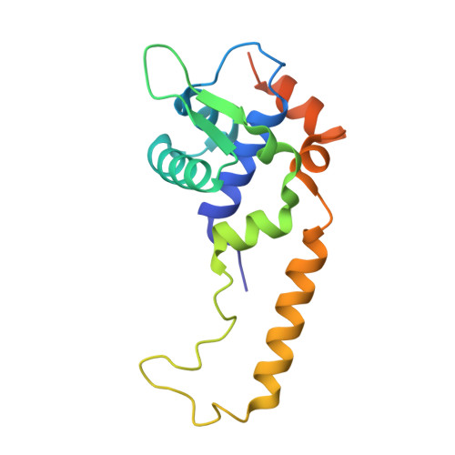
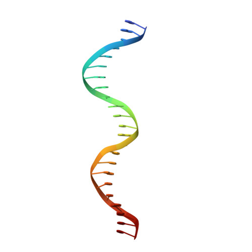
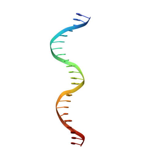
Structural determinants of DNA recognition by the NO sensor NsrR and related Rrf2-type [FeS]-transcription factors.

Rohac, R.Crack, J.C.de Rosny, E.Gigarel, O.Le Brun, N.E.Fontecilla-Camps, J.C.Volbeda, A.

(2022) Commun Biol 5: 769-769

  • DOI: https://doi.org/10.1038/s42003-022-03745-7
  • Primary Citation of Related Structures:  
    7B0C

  • PubMed Abstract: 

    Several transcription factors of the Rrf2 family use an iron-sulfur cluster to regulate DNA binding through effectors such as nitric oxide (NO), cellular redox status and iron levels. [4Fe-4S]-NsrR from Streptomyces coelicolor (ScNsrR) modulates expression of three different genes via reaction and complex formation with variable amounts of NO, which results in detoxification of this gas. Here, we report the crystal structure of ScNsrR complexed with an hmpA1 gene operator fragment and compare it with those previously reported for [2Fe-2S]-RsrR/rsrR and apo-IscR/hyA complexes. Important structural differences reside in the variation of the DNA minor and major groove widths. In addition, different DNA curvatures and different interactions with the protein sensors are observed. We also report studies of NsrR binding to four hmpA1 variants, which indicate that flexibility in the central region is not a key binding determinant. Our study explores the promotor binding specificities of three closely related transcriptional regulators.


  • Organizational Affiliation

    Univ. Grenoble Alpes, CEA, CNRS, Institut de Biologie Structurale, Metalloproteins Unit, F-38000, Grenoble, France.


Macromolecules

Find similar proteins by:  (by identity cutoff)  |  3D Structure
Entity ID: 1
MoleculeChains Sequence LengthOrganismDetailsImage
HTH-type transcriptional repressor NsrR
A, B
161Streptomyces coelicolor A3(2)Mutation(s): 0 
Gene Names: nsrRSCO7427SC6D11.23
UniProt
Find proteins for Q9L132 (Streptomyces coelicolor (strain ATCC BAA-471 / A3(2) / M145))
Explore Q9L132 
Go to UniProtKB:  Q9L132
Entity Groups  
Sequence Clusters30% Identity50% Identity70% Identity90% Identity95% Identity100% Identity
UniProt GroupQ9L132
Sequence Annotations
Expand
  • Reference Sequence

Find similar nucleic acids by:  Sequence   |   3D Structure  

Entity ID: 2
MoleculeChains LengthOrganismImage
DNA (5'-D(P*AP*AP*CP*AP*CP*GP*AP*AP*TP*AP*TP*CP*AP*TP*CP*TP*AP*CP*CP*AP*AP*TP*T)-3')23Streptomyces coelicolor A3(2)
Sequence Annotations
Expand
  • Reference Sequence

Find similar nucleic acids by:  Sequence   |   3D Structure  

Entity ID: 3
MoleculeChains LengthOrganismImage
DNA (5'-D(P*AP*AP*TP*TP*GP*GP*TP*AP*GP*AP*TP*GP*AP*TP*AP*TP*TP*CP*GP*TP*GP*TP*T)-3')23Streptomyces coelicolor A3(2)
Sequence Annotations
Expand
  • Reference Sequence
Experimental Data & Validation

Experimental Data

  • Method: X-RAY DIFFRACTION
  • Resolution: 3.00 Å
  • R-Value Free: 0.236 
  • R-Value Work: 0.219 
  • R-Value Observed: 0.220 
  • Space Group: P 65
Unit Cell:
Length ( Å )Angle ( ˚ )
a = 118.81α = 90
b = 118.81β = 90
c = 89.53γ = 120
Software Package:
Software NamePurpose
PHENIXrefinement
PDB_EXTRACTdata extraction
XDSdata reduction
XDSdata scaling
PHASERphasing

Structure Validation

View Full Validation Report


Entry History & Funding Information

Deposition Data


Funding OrganizationLocationGrant Number
Agence Nationale de la Recherche (ANR)FranceANR-18-CE11-0010

Revision History  (Full details and data files)

  • Version 1.0: 2022-06-01
    Type: Initial release
  • Version 1.1: 2022-08-10
    Changes: Database references, Structure summary
  • Version 1.2: 2024-01-31
    Changes: Data collection, Refinement description