


Cryo-EM structure and dynamics of the green-light absorbing proteorhodopsin
Hirschi, S., Kalbermatter, D., Ucurum, Z., Lemmin, T., Fotiadis, D.(2021) Nature Communications 12: 4107
Experimental Data Snapshot
wwPDB Validation 3D Report Full Report

(2021) Nature Communications 12: 4107
Entity ID: 1 | |||||
|---|---|---|---|---|---|
| Molecule | Chains | Sequence Length | Organism | Details | Image |
| Proteorhodopsin | 233 | uncultured Gammaproteobacteria bacterium | Mutation(s): 0 Membrane Entity: Yes | 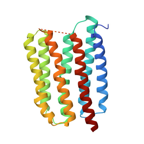 | |
UniProt | |||||
Find proteins for Q6J4G7 (Unknown prokaryotic organism) Explore Q6J4G7 Go to UniProtKB: Q6J4G7 | |||||
Entity Groups | |||||
| Sequence Clusters | 30% Identity50% Identity70% Identity90% Identity95% Identity100% Identity | ||||
| UniProt Group | Q6J4G7 | ||||
Sequence AnnotationsExpand | |||||
| |||||
| Ligands 1 Unique | |||||
|---|---|---|---|---|---|
| ID | Chains | Name / Formula / InChI Key | 2D Diagram | 3D Interactions | |
| RET (Subject of Investigation/LOI) Query on RET | F [auth A], G [auth B], H [auth C], I [auth D], J [auth E] | RETINAL C20 H28 O NCYCYZXNIZJOKI-OVSJKPMPSA-N |  | ||
| Task | Software Package | Version |
|---|---|---|
| RECONSTRUCTION | cryoSPARC | |
| Funding Organization | Location | Grant Number |
|---|---|---|
| Swiss National Science Foundation | Switzerland | -- |
RCSB PDB (citation) is hosted by
RCSB PDB is a member of the

