7AYS

Structure of bovine trypsin determined from single femtosecond snapshots per orientation at room temperature

  • Classification: HYDROLASE
  • Organism(s): Bos taurus
  • Mutation(s): No 

  • Deposited: 2020-11-13 Released: 2021-01-20 
  • Deposition Author(s): Jensen, M.
  • Funding Organization(s): Swedish Research Council

Experimental Data Snapshot

  • Method: X-RAY DIFFRACTION
  • Resolution: 2.10 Å
  • R-Value Free: 0.260 
  • R-Value Work: 0.213 
  • R-Value Observed: 0.215 

wwPDB Validation   3D Report Full Report


Ligand Structure Quality Assessment 


This is version 2.0 of the entry. See complete history


Literature

High-resolution macromolecular crystallography at the FemtoMAX beamline with time-over-threshold photon detection.

Jensen, M.Ahlberg Gagner, V.Cabello Sanchez, J.Bengtsson, A.U.J.Ekstrom, J.C.Bjorg Ulfarsdottir, T.Garcia-Bonete, M.J.Jurgilaitis, A.Kroon, D.Pham, V.T.Checcia, S.Coudert-Alteirac, H.Schewa, S.Rossle, M.Rodilla, H.Stake, J.Zhaunerchyk, V.Larsson, J.Katona, G.

(2021) J Synchrotron Radiat 28: 64-70

  • DOI: https://doi.org/10.1107/S1600577520014599
  • Primary Citation of Related Structures:  
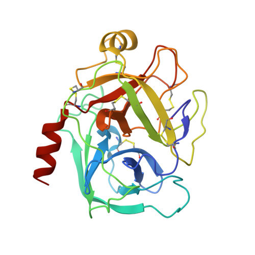
    7AYS

  • PubMed Abstract: 

    Protein dynamics contribute to protein function on different time scales. Ultrafast X-ray diffraction snapshots can visualize the location and amplitude of atom displacements after perturbation. Since amplitudes of ultrafast motions are small, high-quality X-ray diffraction data is necessary for detection. Diffraction from bovine trypsin crystals using single femtosecond X-ray pulses was recorded at FemtoMAX, which is a versatile beamline of the MAX IV synchrotron. The time-over-threshold detection made it possible that single photons are distinguishable even under short-pulse low-repetition-rate conditions. The diffraction data quality from FemtoMAX beamline enables atomic resolution investigation of protein structures. This evaluation is based on the shape of the Wilson plot, cumulative intensity distribution compared with theoretical distribution, I/σ, R merge /R meas and CC 1/2 statistics versus resolution. The FemtoMAX beamline provides an interesting alternative to X-ray free-electron lasers when studying reversible processes in protein crystals.


  • Organizational Affiliation

    Department of Chemistry and Molecular Biology, University of Gothenburg, Gothenburg, Sweden.


Macromolecules
Find similar proteins by:  (by identity cutoff)  |  3D Structure
Entity ID: 1
MoleculeChains Sequence LengthOrganismDetailsImage
Cationic trypsin223Bos taurusMutation(s): 0 
EC: 3.4.21.4
UniProt
Find proteins for P00760 (Bos taurus)
Explore P00760 
Go to UniProtKB:  P00760
Entity Groups  
Sequence Clusters30% Identity50% Identity70% Identity90% Identity95% Identity100% Identity
UniProt GroupP00760
Sequence Annotations
Expand
  • Reference Sequence
Experimental Data & Validation

Experimental Data

  • Method: X-RAY DIFFRACTION
  • Resolution: 2.10 Å
  • R-Value Free: 0.260 
  • R-Value Work: 0.213 
  • R-Value Observed: 0.215 
  • Space Group: P 21 21 21
Unit Cell:
Length ( Å )Angle ( ˚ )
a = 54.84α = 90
b = 58.6β = 90
c = 67.38γ = 90
Software Package:
Software NamePurpose
PHENIXrefinement
XDSdata reduction
XSCALEdata scaling
PHENIXphasing

Structure Validation

View Full Validation Report


Ligand Structure Quality Assessment 


Entry History & Funding Information

Deposition Data

  • Released Date: 2021-01-20 
  • Deposition Author(s): Jensen, M.

Funding OrganizationLocationGrant Number
Swedish Research CouncilSweden2015-06099

Revision History  (Full details and data files)

  • Version 1.0: 2021-01-20
    Type: Initial release
  • Version 2.0: 2021-08-04
    Changes: Atomic model, Author supporting evidence, Data collection, Derived calculations, Non-polymer description, Structure summary