7AUW

Inhibitory complex of human meprin beta with mouse fetuin-B.


Experimental Data Snapshot

  • Method: X-RAY DIFFRACTION
  • Resolution: 2.80 Å
  • R-Value Free: 0.261 
  • R-Value Work: 0.236 
  • R-Value Observed: 0.237 

wwPDB Validation   3D Report Full Report


Ligand Structure Quality Assessment 


This is version 1.1 of the entry. See complete history


Literature

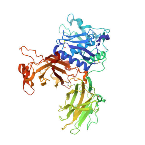
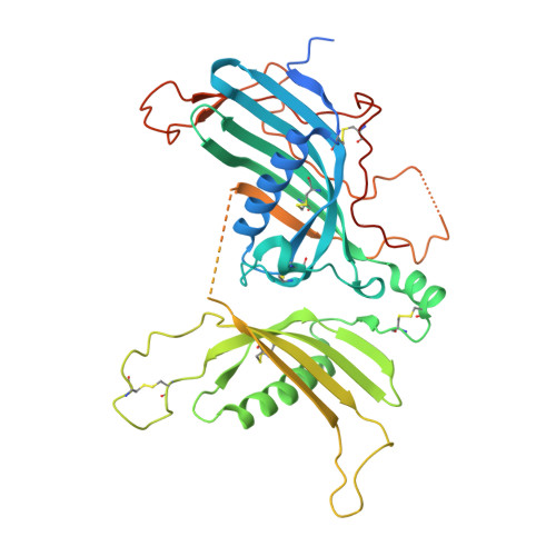
The crystal structure of a 250-kDa heterotetrameric particle explains inhibition of sheddase meprin beta by endogenous fetuin-B.

Eckhard, U.Korschgen, H.von Wiegen, N.Stocker, W.Gomis-Ruth, F.X.

(2021) Proc Natl Acad Sci U S A 118

  • DOI: https://doi.org/10.1073/pnas.2023839118
  • Primary Citation of Related Structures:  
    7AUW

  • PubMed Abstract: 

    Meprin β (Mβ) is a multidomain type-I membrane metallopeptidase that sheds membrane-anchored substrates, releasing their soluble forms. Fetuin-B (FB) is its only known endogenous protein inhibitor. Herein, we analyzed the interaction between the ectodomain of Mβ (MβΔC) and FB, which stabilizes the enzyme and inhibits it with subnanomolar affinity. The MβΔC:FB crystal structure reveals a ∼250-kDa, ∼160-Å polyglycosylated heterotetrameric particle with a remarkable glycan structure. Two FB moieties insert like wedges through a "CPDCP trunk" and two hairpins into the respective peptidase catalytic domains, blocking the catalytic zinc ions through an "aspartate switch" mechanism. Uniquely, the active site clefts are obstructed from subsites S 4 to S 10 ', but S 1 and S 1 ' are spared, which prevents cleavage. Modeling of full-length Mβ reveals an EGF-like domain between MβΔC and the transmembrane segment that likely serves as a hinge to transit between membrane-distal and membrane-proximal conformations for inhibition and catalysis, respectively.


  • Organizational Affiliation

    Proteolysis Laboratory, Department of Structural Biology, Molecular Biology Institute of Barcelona Higher Scientific Research Council, Barcelona Science Park, 08028 Barcelona, Spain.


Macromolecules
Find similar proteins by:  (by identity cutoff)  |  3D Structure
Entity ID: 1
MoleculeChains Sequence LengthOrganismDetailsImage
Meprin A subunit beta
A, C
550Homo sapiensMutation(s): 0 
Gene Names: MEP1B
EC: 3.4.24.63
UniProt & NIH Common Fund Data Resources
Find proteins for Q16820 (Homo sapiens)
Explore Q16820 
Go to UniProtKB:  Q16820
PHAROS:  Q16820
GTEx:  ENSG00000141434 
Entity Groups  
Sequence Clusters30% Identity50% Identity70% Identity90% Identity95% Identity100% Identity
UniProt GroupQ16820
Sequence Annotations
Expand
  • Reference Sequence
Find similar proteins by:  (by identity cutoff)  |  3D Structure
Entity ID: 2
MoleculeChains Sequence LengthOrganismDetailsImage
Fetuin-B
B, D
394Mus musculusMutation(s): 0 
Gene Names: Fetub
UniProt
Find proteins for Q9QXC1 (Mus musculus)
Explore Q9QXC1 
Go to UniProtKB:  Q9QXC1
Entity Groups  
Sequence Clusters30% Identity50% Identity70% Identity90% Identity95% Identity100% Identity
UniProt GroupQ9QXC1
Sequence Annotations
Expand
  • Reference Sequence
Oligosaccharides

Help

Entity ID: 3
MoleculeChains Length2D Diagram Glycosylation3D Interactions
alpha-D-mannopyranose-(1-3)-[alpha-D-mannopyranose-(1-6)]beta-D-mannopyranose-(1-4)-2-acetamido-2-deoxy-beta-D-glucopyranose-(1-4)-[alpha-L-fucopyranose-(1-3)][alpha-L-fucopyranose-(1-6)]2-acetamido-2-deoxy-beta-D-glucopyranose
E, G, M, O
7N-Glycosylation
Glycosylation Resources
GlyTouCan:  G11460AB
GlyCosmos:  G11460AB
GlyGen:  G11460AB
Entity ID: 4
MoleculeChains Length2D Diagram Glycosylation3D Interactions
beta-D-mannopyranose-(1-4)-2-acetamido-2-deoxy-beta-D-glucopyranose-(1-4)-2-acetamido-2-deoxy-beta-D-glucopyranose
F, N, T
3N-Glycosylation
Glycosylation Resources
GlyTouCan:  G15407YE
GlyCosmos:  G15407YE
GlyGen:  G15407YE
Entity ID: 5
MoleculeChains Length2D Diagram Glycosylation3D Interactions
alpha-D-mannopyranose-(1-6)-beta-D-mannopyranose-(1-4)-2-acetamido-2-deoxy-beta-D-glucopyranose-(1-4)-[alpha-L-fucopyranose-(1-3)][alpha-L-fucopyranose-(1-6)]2-acetamido-2-deoxy-beta-D-glucopyranose
H, P
6N-Glycosylation
Glycosylation Resources
GlyTouCan:  G88303DD
GlyCosmos:  G88303DD
GlyGen:  G88303DD
Entity ID: 6
MoleculeChains Length2D Diagram Glycosylation3D Interactions
2-acetamido-2-deoxy-beta-D-glucopyranose-(1-4)-[alpha-L-fucopyranose-(1-6)]2-acetamido-2-deoxy-beta-D-glucopyranose
I, Q
3N-Glycosylation
Glycosylation Resources
GlyTouCan:  G21290RB
GlyCosmos:  G21290RB
GlyGen:  G21290RB
Entity ID: 7
MoleculeChains Length2D Diagram Glycosylation3D Interactions
alpha-D-glucopyranose-(1-3)-alpha-D-mannopyranose-(1-2)-alpha-D-mannopyranose-(1-2)-alpha-D-mannopyranose-(1-3)-[alpha-D-mannopyranose-(1-2)-alpha-D-mannopyranose-(1-3)-[alpha-D-mannopyranose-(1-2)-alpha-D-mannopyranose-(1-6)]alpha-D-mannopyranose-(1-6)]beta-D-mannopyranose-(1-4)-2-acetamido-2-deoxy-beta-D-glucopyranose-(1-4)-2-acetamido-2-deoxy-beta-D-glucopyranose
J, R
12N-Glycosylation
Glycosylation Resources
GlyTouCan:  G19958IL
GlyCosmos:  G19958IL
GlyGen:  G19958IL
Entity ID: 8
MoleculeChains Length2D Diagram Glycosylation3D Interactions
alpha-L-fucopyranose-(1-3)-[alpha-L-fucopyranose-(1-6)]2-acetamido-2-deoxy-beta-D-glucopyranose
K, S
3N-Glycosylation
Glycosylation Resources
GlyTouCan:  G04836KE
GlyCosmos:  G04836KE
GlyGen:  G04836KE
Entity ID: 9
MoleculeChains Length2D Diagram Glycosylation3D Interactions
beta-D-mannopyranose-(1-4)-2-acetamido-2-deoxy-beta-D-glucopyranose-(1-4)-[alpha-L-fucopyranose-(1-3)]2-acetamido-2-deoxy-beta-D-glucopyranose
L
4N-Glycosylation
Glycosylation Resources
GlyTouCan:  G18638YB
GlyCosmos:  G18638YB
GlyGen:  G18638YB
Entity ID: 10
MoleculeChains Length2D Diagram Glycosylation3D Interactions
alpha-L-fucopyranose-(1-3)-[2-acetamido-2-deoxy-beta-D-glucopyranose-(1-4)]2-acetamido-2-deoxy-beta-D-glucopyranose
U
3N-Glycosylation
Glycosylation Resources
GlyTouCan:  G11971MR
GlyCosmos:  G11971MR
GlyGen:  G11971MR
Small Molecules
Ligands 4 Unique
IDChains Name / Formula / InChI Key2D Diagram3D Interactions
NAG
Query on NAG

Download Ideal Coordinates CCD File 
Y [auth B]2-acetamido-2-deoxy-beta-D-glucopyranose
C8 H15 N O6
OVRNDRQMDRJTHS-FMDGEEDCSA-N
SO4
Query on SO4

Download Ideal Coordinates CCD File 
BA [auth C],
X [auth A]
SULFATE ION
O4 S
QAOWNCQODCNURD-UHFFFAOYSA-L
ZN
Query on ZN

Download Ideal Coordinates CCD File 
AA [auth C],
W [auth A]
ZINC ION
Zn
PTFCDOFLOPIGGS-UHFFFAOYSA-N
NA
Query on NA

Download Ideal Coordinates CCD File 
V [auth A],
Z [auth C]
SODIUM ION
Na
FKNQFGJONOIPTF-UHFFFAOYSA-N
Experimental Data & Validation

Experimental Data

  • Method: X-RAY DIFFRACTION
  • Resolution: 2.80 Å
  • R-Value Free: 0.261 
  • R-Value Work: 0.236 
  • R-Value Observed: 0.237 
  • Space Group: P 1 21 1
Unit Cell:
Length ( Å )Angle ( ˚ )
a = 126.66α = 90
b = 88.24β = 113.82
c = 156.66γ = 90
Software Package:
Software NamePurpose
BUSTERrefinement
XDSdata reduction
XSCALEdata scaling
PHENIXphasing

Structure Validation

View Full Validation Report


Ligand Structure Quality Assessment 


Entry History 

Deposition Data

Revision History  (Full details and data files)

  • Version 1.0: 2021-04-14
    Type: Initial release
  • Version 1.1: 2024-01-31
    Changes: Data collection, Database references, Refinement description