7APJ

Structure of autoinhibited Akt1 reveals mechanism of PIP3-mediated activation


Experimental Data Snapshot

  • Method: X-RAY DIFFRACTION
  • Resolution: 2.05 Å
  • R-Value Free: 0.252 
  • R-Value Work: 0.211 
  • R-Value Observed: 0.213 

wwPDB Validation   3D Report Full Report


This is version 1.1 of the entry. See complete history


Literature

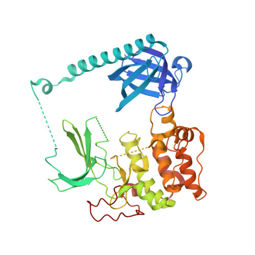
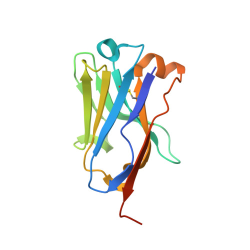
Structure of autoinhibited Akt1 reveals mechanism of PIP 3 -mediated activation.

Truebestein, L.Hornegger, H.Anrather, D.Hartl, M.Fleming, K.D.Stariha, J.T.B.Pardon, E.Steyaert, J.Burke, J.E.Leonard, T.A.

(2021) Proc Natl Acad Sci U S A 118

  • DOI: https://doi.org/10.1073/pnas.2101496118
  • Primary Citation of Related Structures:  
    7APJ

  • PubMed Abstract: 

    The protein kinase Akt is one of the primary effectors of growth factor signaling in the cell. Akt responds specifically to the lipid second messengers phosphatidylinositol-3,4,5-trisphosphate [PI(3,4,5)P 3 ] and phosphatidylinositol-3,4-bisphosphate [PI(3,4)P 2 ] via its PH domain, leading to phosphorylation of its activation loop and the hydrophobic motif of its kinase domain, which are critical for activity. We have now determined the crystal structure of Akt1, revealing an autoinhibitory interface between the PH and kinase domains that is often mutated in cancer and overgrowth disorders. This interface persists even after stoichiometric phosphorylation, thereby restricting maximum Akt activity to PI(3,4,5)P 3 - or PI(3,4)P 2 -containing membranes. Our work helps to resolve the roles of lipids and phosphorylation in the activation of Akt and has wide implications for the spatiotemporal control of Akt and potentially lipid-activated kinase signaling in general.


  • Organizational Affiliation

    Department of Structural and Computational Biology, Max Perutz Labs, Vienna BioCenter, 1030 Vienna, Austria.


Macromolecules
Find similar proteins by:  (by identity cutoff)  |  3D Structure
Entity ID: 1
MoleculeChains Sequence LengthOrganismDetailsImage
RAC-alpha serine/threonine-protein kinase,Non-specific serine/threonine protein kinase,RAC-alpha serine/threonine-protein kinase440Homo sapiensDanio rerioMutation(s): 1 
Gene Names: AKT1PKBRACakt1
EC: 2.7.11.1
Membrane Entity: Yes 
UniProt & NIH Common Fund Data Resources
Find proteins for P31749 (Homo sapiens)
Explore P31749 
Go to UniProtKB:  P31749
PHAROS:  P31749
GTEx:  ENSG00000142208 
Find proteins for M4MD44 (Danio rerio)
Explore M4MD44 
Go to UniProtKB:  M4MD44
Entity Groups  
Sequence Clusters30% Identity50% Identity70% Identity90% Identity95% Identity100% Identity
UniProt GroupsP31749M4MD44
Sequence Annotations
Expand
  • Reference Sequence
Find similar proteins by:  (by identity cutoff)  |  3D Structure
Entity ID: 2
MoleculeChains Sequence LengthOrganismDetailsImage
NB41126Lama glamaMutation(s): 0 
Entity Groups  
Sequence Clusters30% Identity50% Identity70% Identity90% Identity95% Identity100% Identity
Sequence Annotations
Expand
  • Reference Sequence
Experimental Data & Validation

Experimental Data

  • Method: X-RAY DIFFRACTION
  • Resolution: 2.05 Å
  • R-Value Free: 0.252 
  • R-Value Work: 0.211 
  • R-Value Observed: 0.213 
  • Space Group: P 21 21 21
Unit Cell:
Length ( Å )Angle ( ˚ )
a = 70.008α = 90
b = 72.197β = 90
c = 120.173γ = 90
Software Package:
Software NamePurpose
PHENIXrefinement
PHENIXrefinement
XDSdata reduction
Aimlessdata scaling
PHASERphasing

Structure Validation

View Full Validation Report


Entry History & Funding Information

Deposition Data


Funding OrganizationLocationGrant Number
Austrian Science FundAustriaP28135
Austrian Science FundAustriaP30584
Austrian Science FundAustriaP33066
Austrian Science FundAustriaT915

Revision History  (Full details and data files)

  • Version 1.0: 2021-08-25
    Type: Initial release
  • Version 1.1: 2024-01-31
    Changes: Data collection, Refinement description