7AEO

Human ARTD2 in complex with DNA oligonucleotides


Experimental Data Snapshot

  • Method: X-RAY DIFFRACTION
  • Resolution: 2.80 Å
  • R-Value Free: 0.271 
  • R-Value Work: 0.227 
  • R-Value Observed: 0.229 

wwPDB Validation   3D Report Full Report


This is version 1.3 of the entry. See complete history


Literature

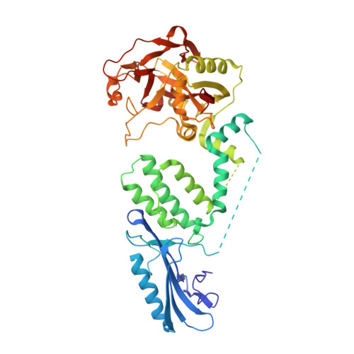
Activation of PARP2/ARTD2 by DNA damage induces conformational changes relieving enzyme autoinhibition.

Obaji, E.Maksimainen, M.M.Galera-Prat, A.Lehtio, L.

(2021) Nat Commun 12: 3479-3479

  • DOI: https://doi.org/10.1038/s41467-021-23800-x
  • Primary Citation of Related Structures:  
    7AEO

  • PubMed Abstract: 

    Human PARP2/ARTD2 is an ADP-ribosyltransferase which, when activated by 5'-phosphorylated DNA ends, catalyses poly-ADP-ribosylation of itself, other proteins and DNA. In this study, a crystal structure of PARP2 in complex with an activating 5'-phosphorylated DNA shows that the WGR domain bridges the dsDNA gap and joins the DNA ends. This DNA binding results in major conformational changes, including reorganization of helical fragments, in the PARP2 regulatory domain. A comparison of PARP1 and PARP2 crystal structures reveals how binding to a DNA damage site leads to formation of a catalytically competent conformation. In this conformation, PARP2 is capable of binding substrate NAD + and histone PARylation factor 1 that changes PARP2 residue specificity from glutamate to serine when initiating DNA repair processes. The structure also reveals how the conformational changes in the autoinhibitory regulatory domain would promote the flexibility needed by the enzyme to reach the target macromolecule for ADP-ribosylation.


  • Organizational Affiliation

    Faculty of Biochemistry and Molecular Medicine & Biocenter Oulu, University of Oulu, Oulu, Finland.


Macromolecules

Find similar proteins by:  (by identity cutoff)  |  3D Structure
Entity ID: 1
MoleculeChains Sequence LengthOrganismDetailsImage
Poly [ADP-ribose] polymerase 2496Homo sapiensMutation(s): 0 
Gene Names: PARP2ADPRT2ADPRTL2
EC: 2.4.2.30 (PDB Primary Data), 2.4.2 (PDB Primary Data)
UniProt & NIH Common Fund Data Resources
Find proteins for Q9UGN5 (Homo sapiens)
Explore Q9UGN5 
Go to UniProtKB:  Q9UGN5
PHAROS:  Q9UGN5
GTEx:  ENSG00000129484 
Entity Groups  
Sequence Clusters30% Identity50% Identity70% Identity90% Identity95% Identity100% Identity
UniProt GroupQ9UGN5
Sequence Annotations
Expand
  • Reference Sequence

Find similar nucleic acids by:  Sequence   |   3D Structure  

Entity ID: 2
MoleculeChains LengthOrganismImage
DNA16synthetic construct
Sequence Annotations
Expand
  • Reference Sequence

Find similar nucleic acids by:  Sequence   |   3D Structure  

Entity ID: 3
MoleculeChains LengthOrganismImage
DNA16synthetic construct
Sequence Annotations
Expand
  • Reference Sequence
Experimental Data & Validation

Experimental Data

  • Method: X-RAY DIFFRACTION
  • Resolution: 2.80 Å
  • R-Value Free: 0.271 
  • R-Value Work: 0.227 
  • R-Value Observed: 0.229 
  • Space Group: I 41 2 2
Unit Cell:
Length ( Å )Angle ( ˚ )
a = 166.83α = 90
b = 166.83β = 90
c = 143.84γ = 90
Software Package:
Software NamePurpose
PHENIXrefinement
XDSdata reduction
XSCALEdata scaling
MrBUMPphasing

Structure Validation

View Full Validation Report


Entry History & Funding Information

Deposition Data


Funding OrganizationLocationGrant Number
Jane and Aatos Erkko FoundationFinland--
Academy of FinlandFinland287063
Academy of FinlandFinland294085
Academy of FinlandFinland319299

Revision History  (Full details and data files)

  • Version 1.0: 2021-05-19
    Type: Initial release
  • Version 1.1: 2021-05-26
    Changes: Database references, Source and taxonomy, Structure summary
  • Version 1.2: 2021-06-23
    Changes: Database references
  • Version 1.3: 2024-01-31
    Changes: Data collection, Database references, Refinement description