7AEJ

Crystal structure of asymmetric HIV-1 gp41 containing all membrane anchors


Experimental Data Snapshot

  • Method: X-RAY DIFFRACTION
  • Resolution: 3.80 Å
  • R-Value Free: 0.308 
  • R-Value Work: 0.265 
  • R-Value Observed: 0.268 

wwPDB Validation   3D Report Full Report


This is version 1.1 of the entry. See complete history


Literature

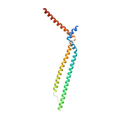
Structure of HIV-1 gp41 with its membrane anchors targeted by neutralizing antibodies.

Caillat, C.Guilligay, D.Torralba, J.Friedrich, N.Nieva, J.L.Trkola, A.Chipot, C.J.Dehez, F.L.Weissenhorn, W.

(2021) Elife 10

  • DOI: https://doi.org/10.7554/eLife.65005
  • Primary Citation of Related Structures:  
    7AEJ

  • PubMed Abstract: 

    The HIV-1 gp120/gp41 trimer undergoes a series of conformational changes in order to catalyze gp41-induced fusion of viral and cellular membranes. Here, we present the crystal structure of gp41 locked in a fusion intermediate state by an MPER-specific neutralizing antibody. The structure illustrates the conformational plasticity of the six membrane anchors arranged asymmetrically with the fusion peptides and the transmembrane regions pointing into different directions. Hinge regions located adjacent to the fusion peptide and the transmembrane region facilitate the conformational flexibility that allows high-affinity binding of broadly neutralizing anti-MPER antibodies. Molecular dynamics simulation of the MPER Ab-stabilized gp41 conformation reveals a possible transition pathway into the final post-fusion conformation with the central fusion peptides forming a hydrophobic core with flanking transmembrane regions. This suggests that MPER-specific broadly neutralizing antibodies can block final steps of refolding of the fusion peptide and the transmembrane region, which is required for completing membrane fusion.


  • Organizational Affiliation

    Univ. Grenoble Alpes, CEA, CNRS, Institut de Biologie Structurale (IBS), Grenoble, France.


Macromolecules
Find similar proteins by:  (by identity cutoff)  |  3D Structure
Entity ID: 1
MoleculeChains Sequence LengthOrganismDetailsImage
Envelope glycoprotein gp160,Envelope glycoprotein gp160A [auth C],
B [auth A],
C [auth B]
192Human immunodeficiency virus 1Mutation(s): 2 
Gene Names: env
Membrane Entity: Yes 
UniProt
Find proteins for P04578 (Human immunodeficiency virus type 1 group M subtype B (isolate HXB2))
Explore P04578 
Go to UniProtKB:  P04578
Entity Groups  
Sequence Clusters30% Identity50% Identity70% Identity90% Identity95% Identity100% Identity
UniProt GroupP04578
Sequence Annotations
Expand
  • Reference Sequence
Find similar proteins by:  (by identity cutoff)  |  3D Structure
Entity ID: 2
MoleculeChains Sequence LengthOrganismDetailsImage
2H10122Lama glamaMutation(s): 0 
Entity Groups  
Sequence Clusters30% Identity50% Identity70% Identity90% Identity95% Identity100% Identity
Sequence Annotations
Expand
  • Reference Sequence
Experimental Data & Validation

Experimental Data

  • Method: X-RAY DIFFRACTION
  • Resolution: 3.80 Å
  • R-Value Free: 0.308 
  • R-Value Work: 0.265 
  • R-Value Observed: 0.268 
  • Space Group: C 2 2 21
Unit Cell:
Length ( Å )Angle ( ˚ )
a = 96.75α = 90
b = 101.414β = 90
c = 234.417γ = 90
Software Package:
Software NamePurpose
PHENIXrefinement
XDSdata reduction
Aimlessdata scaling
STARANISOdata scaling
PHENIXphasing

Structure Validation

View Full Validation Report


Entry History 

Deposition Data

Revision History  (Full details and data files)

  • Version 1.0: 2021-05-26
    Type: Initial release
  • Version 1.1: 2024-01-31
    Changes: Data collection, Database references, Refinement description