


SFX structure of dehaloperoxidase B from Amphitrite ornata in the oxyferrous form
Moreno Chicano, T., Ebrahim, A., Worrall, J.W., Axford, D.A., Owada, S., Tosha, T., Sugimoto, H., Strange, R.W., Owen, R.L., Hough, M.A.To be published.
Experimental Data Snapshot
Entity ID: 1 | |||||
|---|---|---|---|---|---|
| Molecule | Chains | Sequence Length | Organism | Details | Image |
| Dehaloperoxidase B | 138 | Amphitrite ornata | Mutation(s): 0 | 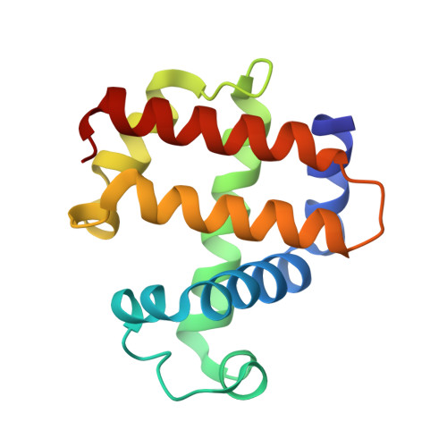 | |
UniProt | |||||
Find proteins for Q9NAV7 (Amphitrite ornata) Explore Q9NAV7 Go to UniProtKB: Q9NAV7 | |||||
Entity Groups | |||||
| Sequence Clusters | 30% Identity50% Identity70% Identity90% Identity95% Identity100% Identity | ||||
| UniProt Group | Q9NAV7 | ||||
Sequence AnnotationsExpand | |||||
| |||||
| Ligands 3 Unique | |||||
|---|---|---|---|---|---|
| ID | Chains | Name / Formula / InChI Key | 2D Diagram | 3D Interactions | |
| HEM Query on HEM | C [auth A], F [auth B] | PROTOPORPHYRIN IX CONTAINING FE C34 H32 Fe N4 O4 KABFMIBPWCXCRK-RGGAHWMASA-L |  | ||
| SO4 Query on SO4 | D [auth A], E [auth A] | SULFATE ION O4 S QAOWNCQODCNURD-UHFFFAOYSA-L |  | ||
| OXY Query on OXY | G [auth B] | OXYGEN MOLECULE O2 MYMOFIZGZYHOMD-UHFFFAOYSA-N |  | ||
| Length ( Å ) | Angle ( ˚ ) |
|---|---|
| a = 61.31 | α = 90 |
| b = 68.1 | β = 90 |
| c = 68.33 | γ = 90 |
| Software Name | Purpose |
|---|---|
| REFMAC | refinement |
| PDB_EXTRACT | data extraction |
| CrystFEL | data reduction |
| CrystFEL | data scaling |
| REFMAC | phasing |
| Funding Organization | Location | Grant Number |
|---|---|---|
| Biotechnology and Biological Sciences Research Council (BBSRC) | United Kingdom | BB/R021015/1 |
RCSB PDB (citation) is hosted by
RCSB PDB is a member of the

