7AAL

Crystal structure of the F-BAR domain of PSTIPIP1, G258A mutant


Experimental Data Snapshot

  • Method: X-RAY DIFFRACTION
  • Resolution: 1.97 Å
  • R-Value Free: 0.219 
  • R-Value Work: 0.186 
  • R-Value Observed: 0.188 

wwPDB Validation   3D Report Full Report


This is version 1.3 of the entry. See complete history


Literature

PSTPIP1-LYP phosphatase interaction: structural basis and implications for autoinflammatory disorders.

Manso, J.A.Marcos, T.Ruiz-Martin, V.Casas, J.Alcon, P.Sanchez Crespo, M.Bayon, Y.de Pereda, J.M.Alonso, A.

(2022) Cell Mol Life Sci 79: 131-131

  • DOI: https://doi.org/10.1007/s00018-022-04173-w
  • Primary Citation of Related Structures:  
    7AAL, 7AAM, 7AAN

  • PubMed Abstract: 

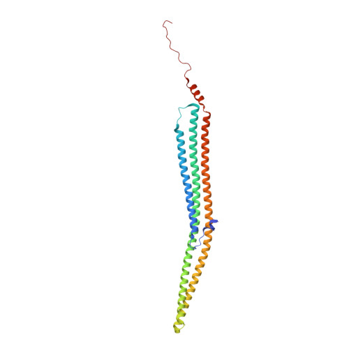
    Mutations in the adaptor protein PSTPIP1 cause a spectrum of autoinflammatory diseases, including PAPA and PAMI; however, the mechanism underlying these diseases remains unknown. Most of these mutations lie in PSTPIP1 F-BAR domain, which binds to LYP, a protein tyrosine phosphatase associated with arthritis and lupus. To shed light on the mechanism by which these mutations generate autoinflammatory disorders, we solved the structure of the F-BAR domain of PSTPIP1 alone and bound to the C-terminal homology segment of LYP, revealing a novel mechanism of recognition of Pro-rich motifs by proteins in which a single LYP molecule binds to the PSTPIP1 F-BAR dimer. The residues R228, D246, E250, and E257 of PSTPIP1 that are mutated in immunological diseases directly interact with LYP. These findings link the disruption of the PSTPIP1/LYP interaction to these diseases, and support a critical role for LYP phosphatase in their pathogenesis.


  • Organizational Affiliation

    Instituto de Biología Molecular y Celular del Cáncer (IBMCC), CSIC-Universidad de Salamanca, Campus Unamuno, 37007, Salamanca, Spain.


Macromolecules
Find similar proteins by:  (by identity cutoff)  |  3D Structure
Entity ID: 1
MoleculeChains Sequence LengthOrganismDetailsImage
Proline-serine-threonine phosphatase-interacting protein 1
A, B
292Homo sapiensMutation(s): 1 
Gene Names: PSTPIP1CD2BP1
Membrane Entity: Yes 
UniProt & NIH Common Fund Data Resources
Find proteins for O43586 (Homo sapiens)
Explore O43586 
Go to UniProtKB:  O43586
PHAROS:  O43586
GTEx:  ENSG00000140368 
Entity Groups  
Sequence Clusters30% Identity50% Identity70% Identity90% Identity95% Identity100% Identity
UniProt GroupO43586
Sequence Annotations
Expand
  • Reference Sequence
Experimental Data & Validation

Experimental Data

  • Method: X-RAY DIFFRACTION
  • Resolution: 1.97 Å
  • R-Value Free: 0.219 
  • R-Value Work: 0.186 
  • R-Value Observed: 0.188 
  • Space Group: P 21 21 21
Unit Cell:
Length ( Å )Angle ( ˚ )
a = 48.191α = 90
b = 73.023β = 90
c = 205.248γ = 90
Software Package:
Software NamePurpose
XDSdata reduction
Aimlessdata scaling
STARANISOdata scaling
PHASERphasing
PHENIXrefinement

Structure Validation

View Full Validation Report


Entry History & Funding Information

Deposition Data


Funding OrganizationLocationGrant Number
Other privateSpain--

Revision History  (Full details and data files)

  • Version 1.0: 2022-02-23
    Type: Initial release
  • Version 1.1: 2022-03-02
    Changes: Database references
  • Version 1.2: 2023-02-08
    Changes: Structure summary
  • Version 1.3: 2024-02-07
    Changes: Data collection, Refinement description