7AAK

Human porphobilinogen deaminase R173W mutant crystallized in the ES2 intermediate state


Experimental Data Snapshot

  • Method: X-RAY DIFFRACTION
  • Resolution: 1.70 Å
  • R-Value Free: 0.213 
  • R-Value Work: 0.183 
  • R-Value Observed: 0.184 

wwPDB Validation   3D Report Full Report


Ligand Structure Quality Assessment 


This is version 1.1 of the entry. See complete history


Literature

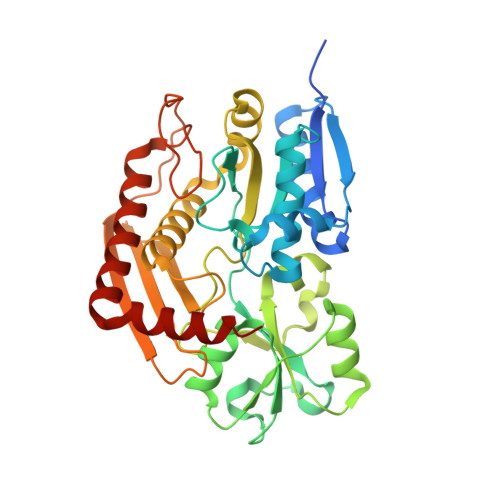
Characterization of porphobilinogen deaminase mutants reveals that arginine-173 is crucial for polypyrrole elongation mechanism.

Bustad, H.J.Kallio, J.P.Laitaoja, M.Toska, K.Kursula, I.Martinez, A.Janis, J.

(2021) iScience 24: 102152-102152

  • DOI: https://doi.org/10.1016/j.isci.2021.102152
  • Primary Citation of Related Structures:  
    7AAJ, 7AAK

  • PubMed Abstract: 

    Porphobilinogen deaminase (PBGD), the third enzyme in the heme biosynthesis, catalyzes the sequential coupling of four porphobilinogen (PBG) molecules into a heme precursor. Mutations in PBGD are associated with acute intermittent porphyria (AIP), a rare metabolic disorder. We used Fourier transform ion cyclotron resonance mass spectrometry (FT-ICR MS) to demonstrate that wild-type PBGD and AIP-associated mutant R167W both existed as holoenzymes (E holo ) covalently attached to the dipyrromethane cofactor, and three intermediate complexes, ES, ES 2 , and ES 3 , where S represents PBG. In contrast, only ES 2 was detected in AIP-associated mutant R173W, indicating that the formation of ES 3 is inhibited. The R173W crystal structure in the ES 2 -state revealed major rearrangements of the loops around the active site, compared to wild-type PBGD in the E holo -state. These results contribute to elucidating the structural pathogenesis of two common AIP-associated mutations and reveal the important structural role of Arg173 in the polypyrrole elongation mechanism.


  • Organizational Affiliation

    Department of Biomedicine, University of Bergen, Jonas Lies vei 91, 5009 Bergen, Norway.


Macromolecules
Find similar proteins by:  (by identity cutoff)  |  3D Structure
Entity ID: 1
MoleculeChains Sequence LengthOrganismDetailsImage
Porphobilinogen deaminase
A, B
363Homo sapiensMutation(s): 1 
Gene Names: HMBSPBGDUPS
EC: 2.5.1.61
UniProt & NIH Common Fund Data Resources
Find proteins for P08397 (Homo sapiens)
Explore P08397 
Go to UniProtKB:  P08397
PHAROS:  P08397
GTEx:  ENSG00000256269 
Entity Groups  
Sequence Clusters30% Identity50% Identity70% Identity90% Identity95% Identity100% Identity
UniProt GroupP08397
Sequence Annotations
Expand
  • Reference Sequence
Small Molecules
Ligands 2 Unique
IDChains Name / Formula / InChI Key2D Diagram3D Interactions
7J8 (Subject of Investigation/LOI)
Query on 7J8

Download Ideal Coordinates CCD File 
C [auth A],
F [auth B]
3-[4-(2-hydroxy-2-oxoethyl)-5-[[4-(2-hydroxy-2-oxoethyl)-5-[[4-(2-hydroxy-2-oxoethyl)-5-[[4-(2-hydroxy-2-oxoethyl)-3-(3-hydroxy-3-oxopropyl)-5-methyl-1~{H}-pyrrol-2-yl]methyl]-3-(3-hydroxy-3-oxopropyl)-1~{H}-pyrrol-2-yl]methyl]-3-(3-hydroxy-3-oxopropyl)-1~{H}-pyrrol-2-yl]methyl]-1~{H}-pyrrol-3-yl]propanoic acid
C40 H46 N4 O16
VHZWMEJYGVBAFI-UHFFFAOYSA-N
GOL
Query on GOL

Download Ideal Coordinates CCD File 
D [auth A],
E [auth A],
G [auth B],
H [auth B]
GLYCEROL
C3 H8 O3
PEDCQBHIVMGVHV-UHFFFAOYSA-N
Experimental Data & Validation

Experimental Data

  • Method: X-RAY DIFFRACTION
  • Resolution: 1.70 Å
  • R-Value Free: 0.213 
  • R-Value Work: 0.183 
  • R-Value Observed: 0.184 
  • Space Group: P 21 21 21
Unit Cell:
Length ( Å )Angle ( ˚ )
a = 81.241α = 90
b = 86.127β = 90
c = 107.39γ = 90
Software Package:
Software NamePurpose
XDSdata reduction
Aimlessdata scaling
PHASERphasing
PHENIXrefinement

Structure Validation

View Full Validation Report


Ligand Structure Quality Assessment 


Entry History 

Deposition Data

Revision History  (Full details and data files)

  • Version 1.0: 2021-02-17
    Type: Initial release
  • Version 1.1: 2021-03-17
    Changes: Database references