Structure-functional relationship of cellular retinoic acid-binding proteins I and II interacting with natural and synthetic ligands.
Tomlinson, C.W.E., Cornish, K.A.S., Whiting, A., Pohl, E.(2021) Acta Crystallogr D Struct Biol 77: 164-175
- PubMed: 33559606
- DOI: https://doi.org/10.1107/S2059798320015247
- Primary Citation of Related Structures:
7A9Y, 7A9Z, 7AA0, 7AA1 - PubMed Abstract:
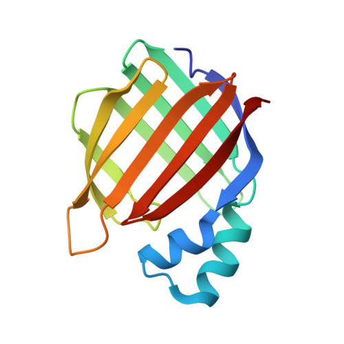
A detailed understanding of the interactions between small-molecule ligands and their proposed binding targets is of the utmost importance for modern drug-development programs. Cellular retinoic acid-binding proteins I and II (CRABPI and CRABPII) facilitate a number of vital retinoid signalling pathways in mammalian cells and offer a gateway to manipulation of signalling that could potentially reduce phenotypes in serious diseases, including cancer and neurodegeneration. Although structurally very similar, the two proteins possess distinctly different biological functions, with their signalling influence being exerted through both genomic and nongenomic pathways. In this article, crystal structures are presented of the L29C mutant of Homo sapiens CRABPI in complex with naturally occurring fatty acids (1.64 Å resolution) and with the synthetic retinoid DC645 (2.41 Å resolution), and of CRABPII in complex with the ligands DC479 (1.80 Å resolution) and DC645 (1.71 Å resolution). DC645 and DC479 are two potential drug compounds identified in a recent synthetic retinoid development program. In particular, DC645 has recently been shown to have disease-modifying capabilities in neurodegenerative disease models by activating both genomic and nongenomic signalling pathways. These co-crystal structures demonstrate a canonical binding behaviour akin to that exhibited with all-trans-retinoic acid and help to explain how the compounds are able to exert an influence on part of the retinoid signalling cascade.
Organizational Affiliation:
Department of Chemistry, Durham University, Lower Mountjoy, South Road, Durham DH1 3LE, United Kingdom.