7A5E

OSM-3 kinesin motor domain complexed with Mg.AMPPNP


Experimental Data Snapshot

  • Method: X-RAY DIFFRACTION
  • Resolution: 1.90 Å
  • R-Value Free: 0.231 
  • R-Value Work: 0.206 
  • R-Value Observed: 0.208 

wwPDB Validation   3D Report Full Report


Ligand Structure Quality Assessment 


This is version 1.3 of the entry. See complete history


Literature

Structural snapshots of the kinesin-2 OSM-3 along its nucleotide cycle: implications for the ATP hydrolysis mechanism.

Varela, P.F.Chenon, M.Velours, C.Verhey, K.J.Menetrey, J.Gigant, B.

(2021) FEBS Open Bio 11: 564-577

  • DOI: https://doi.org/10.1002/2211-5463.13101
  • Primary Citation of Related Structures:  
    7A3Z, 7A40, 7A5E

  • PubMed Abstract: 

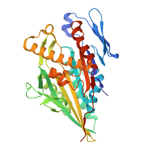
    Motile kinesins are motor proteins that translocate along microtubules as they hydrolyze ATP. They share a conserved motor domain which harbors both ATPase and microtubule-binding activities. An ATP hydrolysis mechanism involving two water molecules has been proposed based on the structure of the kinesin-5 Eg5 bound to an ATP analog. Whether this mechanism is general in the kinesin superfamily remains uncertain. Here, we present structural snapshots of the motor domain of OSM-3 along its nucleotide cycle. OSM-3 belongs to the homodimeric kinesin-2 subfamily and is the Caenorhabditis elegans homologue of human KIF17. OSM-3 bound to ADP or devoid of a nucleotide shows features of ADP-kinesins with a docked neck linker. When bound to an ATP analog, OSM-3 adopts a conformation similar to those of several ATP-like kinesins, either isolated or bound to tubulin. Moreover, the OSM-3 nucleotide-binding site is virtually identical to that of ATP-like Eg5, demonstrating a shared ATPase mechanism. Therefore, our data extend to kinesin-2 the two-water ATP hydrolysis mechanism and further suggest that it is universal within the kinesin superfamily. PROTEIN DATABASE ENTRIES: 7A3Z, 7A40, 7A5E.


  • Organizational Affiliation

    Université Paris-Saclay, CEA, CNRS, Institute for Integrative Biology of the Cell (I2BC), Gif-sur-Yvette, France.


Macromolecules
Find similar proteins by:  (by identity cutoff)  |  3D Structure
Entity ID: 1
MoleculeChains Sequence LengthOrganismDetailsImage
Osmotic avoidance abnormal protein 3
A, B
346Caenorhabditis elegansMutation(s): 0 
Gene Names: osm-3M02B7.3
UniProt
Find proteins for P46873 (Caenorhabditis elegans)
Explore P46873 
Go to UniProtKB:  P46873
Entity Groups  
Sequence Clusters30% Identity50% Identity70% Identity90% Identity95% Identity100% Identity
UniProt GroupP46873
Sequence Annotations
Expand
  • Reference Sequence
Small Molecules
Ligands 4 Unique
IDChains Name / Formula / InChI Key2D Diagram3D Interactions
ANP
Query on ANP

Download Ideal Coordinates CCD File 
C [auth A],
J [auth B]
PHOSPHOAMINOPHOSPHONIC ACID-ADENYLATE ESTER
C10 H17 N6 O12 P3
PVKSNHVPLWYQGJ-KQYNXXCUSA-N
SO4
Query on SO4

Download Ideal Coordinates CCD File 
E [auth A],
F [auth A],
L [auth B],
M [auth B]
SULFATE ION
O4 S
QAOWNCQODCNURD-UHFFFAOYSA-L
GOL
Query on GOL

Download Ideal Coordinates CCD File 
G [auth A]
H [auth A]
I [auth A]
N [auth B]
O [auth B]
G [auth A],
H [auth A],
I [auth A],
N [auth B],
O [auth B],
P [auth B],
Q [auth B]
GLYCEROL
C3 H8 O3
PEDCQBHIVMGVHV-UHFFFAOYSA-N
MG
Query on MG

Download Ideal Coordinates CCD File 
D [auth A],
K [auth B]
MAGNESIUM ION
Mg
JLVVSXFLKOJNIY-UHFFFAOYSA-N
Experimental Data & Validation

Experimental Data

  • Method: X-RAY DIFFRACTION
  • Resolution: 1.90 Å
  • R-Value Free: 0.231 
  • R-Value Work: 0.206 
  • R-Value Observed: 0.208 
  • Space Group: C 2 2 21
Unit Cell:
Length ( Å )Angle ( ˚ )
a = 100.03α = 90
b = 102.2β = 90
c = 140.95γ = 90
Software Package:
Software NamePurpose
BUSTERrefinement
XDSdata reduction
XDSdata scaling
PHASERphasing

Structure Validation

View Full Validation Report


Ligand Structure Quality Assessment 


Entry History 

Deposition Data

Revision History  (Full details and data files)

  • Version 1.0: 2021-02-03
    Type: Initial release
  • Version 1.1: 2021-02-10
    Changes: Database references
  • Version 1.2: 2021-03-17
    Changes: Database references
  • Version 1.3: 2024-01-31
    Changes: Data collection, Database references, Refinement description