NMR structure of the Vibrio vulnificus ribosomal protein S1 domains D3 and D4 provides insights into molecular recognition of single-stranded RNAs.
Qureshi, N.S., Matzel, T., Cetiner, E.C., Schnieders, R., Jonker, H.R.A., Schwalbe, H., Furtig, B.(2021) Nucleic Acids Res 49: 7753-7764
- PubMed: 34223902
- DOI: https://doi.org/10.1093/nar/gkab562
- Primary Citation of Related Structures:
7A05 - PubMed Abstract:
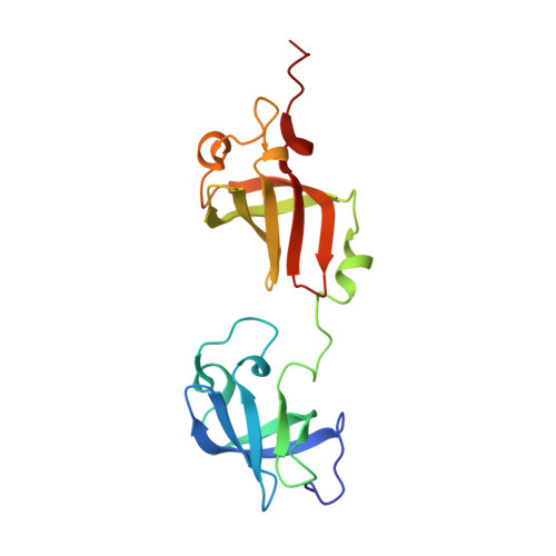
The ribosomal S1 protein (rS1) is indispensable for translation initiation in Gram-negative bacteria. rS1 is a multidomain protein that acts as an RNA chaperone and ensures that mRNAs can bind the ribosome in a single-stranded conformation, which could be related to fast recognition. Although many ribosome structures were solved in recent years, a high-resolution structure of a two-domain mRNA-binding competent rS1 construct is not yet available. Here, we present the NMR solution structure of the minimal mRNA-binding fragment of Vibrio Vulnificus rS1 containing the domains D3 and D4. Both domains are homologues and adapt an oligonucleotide-binding fold (OB fold) motif. NMR titration experiments reveal that recognition of miscellaneous mRNAs occurs via a continuous interaction surface to one side of these structurally linked domains. Using a novel paramagnetic relaxation enhancement (PRE) approach and exploring different spin-labeling positions within RNA, we were able to track the location and determine the orientation of the RNA in the rS1-D34 bound form. Our investigations show that paramagnetically labeled RNAs, spiked into unmodified RNA, can be used as a molecular ruler to provide structural information on protein-RNA complexes. The dynamic interaction occurs on a defined binding groove spanning both domains with identical β2-β3-β5 interfaces. Evidently, the 3'-ends of the cis-acting RNAs are positioned in the direction of the N-terminus of the rS1 protein, thus towards the 30S binding site and adopt a conformation required for translation initiation.
Organizational Affiliation:
Institute for Organic Chemistry and Chemical Biology, Center for Biomolecular Magnetic Resonance (BMRZ), Johann Wolfgang Goethe-Universität, Frankfurt am Main, Hesse 60438, Germany.