7ZXW

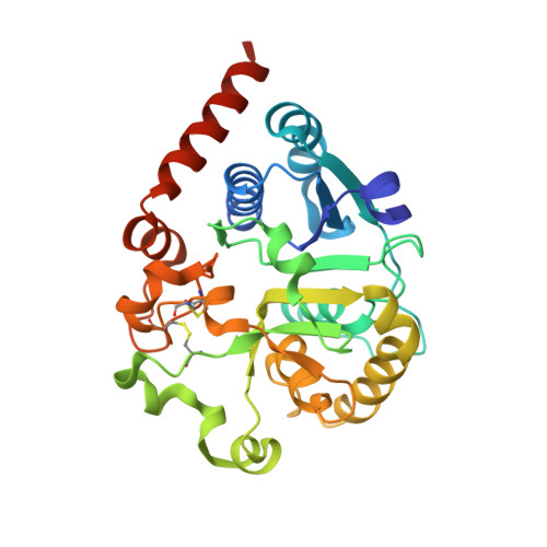
Catalytic domain of UDP-Glucose Glycoprotein Glucosyltransferase from Chaetomium thermophilum in complex with the 5-[(morpholin-4-yl)methyl]quinolin-8-ol inhibitor


Experimental Data Snapshot

  • Method: X-RAY DIFFRACTION
  • Resolution: 2.25 Å
  • R-Value Free: 0.240 
  • R-Value Work: 0.203 
  • R-Value Observed: 0.205 

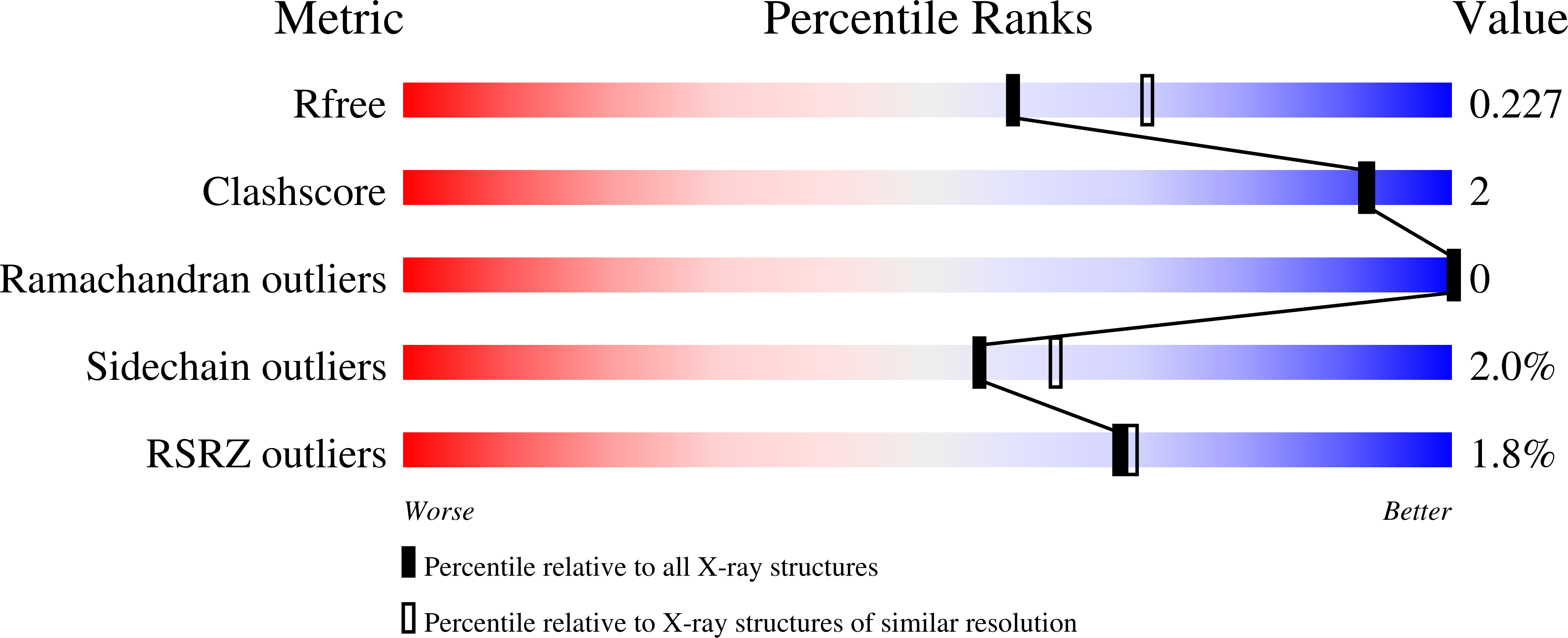
wwPDB Validation   3D Report Full Report


Ligand Structure Quality Assessment 


This is version 1.3 of the entry. See complete history


Literature

Crystal polymorphism in fragment-based lead discovery of ligands of the catalytic domain of UGGT, the glycoprotein folding quality control checkpoint.

Caputo, A.T.Ibba, R.Le Cornu, J.D.Darlot, B.Hensen, M.Lipp, C.B.Marciano, G.Vasiljevic, S.Zitzmann, N.Roversi, P.

(2022) Front Mol Biosci 9: 960248-960248

  • DOI: https://doi.org/10.3389/fmolb.2022.960248
  • Primary Citation of Related Structures:  
    7ZXW

  • PubMed Abstract: 

    None of the current data processing pipelines for X-ray crystallography fragment-based lead discovery (FBLD) consults all the information available when deciding on the lattice and symmetry (i.e., the polymorph) of each soaked crystal. Often, X-ray crystallography FBLD pipelines either choose the polymorph based on cell volume and point-group symmetry of the X-ray diffraction data or leave polymorph attribution to manual intervention on the part of the user. Thus, when the FBLD crystals belong to more than one crystal polymorph, the discovery pipeline can be plagued by space group ambiguity, especially if the polymorphs at hand are variations of the same lattice and, therefore, difficult to tell apart from their morphology and/or their apparent crystal lattices and point groups. In the course of a fragment-based lead discovery effort aimed at finding ligands of the catalytic domain of UDP-glucose glycoprotein glucosyltransferase (UGGT), we encountered a mixture of trigonal crystals and pseudotrigonal triclinic crystals-with the two lattices closely related. In order to resolve that polymorphism ambiguity, we have written and described here a series of Unix shell scripts called CoALLA ( c rystal p o lymorph a nd l igand l ikelihood-based a ssignment). The CoALLA scripts are written in Unix shell and use autoPROC for data processing, CCP4-Dimple / REFMAC5 and BUSTER for refinement, and RHOFIT for ligand docking. The choice of the polymorph is effected by carrying out (in each of the known polymorphs) the tasks of diffraction data indexing, integration, scaling, and structural refinement. The most likely polymorph is then chosen as the one with the best structure refinement R free statistic. The CoALLA scripts further implement a likelihood-based ligand assignment strategy, starting with macromolecular refinement and automated water addition, followed by removal of the water molecules that appear to be fitting ligand density, and a final round of refinement after random perturbation of the refined macromolecular model, in order to obtain unbiased difference density maps for automated ligand placement. We illustrate the use of CoALLA to discriminate between H3 and P1 crystals used for an FBLD effort to find fragments binding to the catalytic domain of Chaetomium thermophilum UGGT.


  • Organizational Affiliation

    Biochemistry Department, Oxford Glycobiology Institute, University of Oxford, Oxford, United Kingdom.


Macromolecules
Find similar proteins by:  (by identity cutoff)  |  3D Structure
Entity ID: 1
MoleculeChains Sequence LengthOrganismDetailsImage
UDP-glucose-glycoprotein glucosyltransferase-like protein295Thermochaetoides thermophila DSM 1495Mutation(s): 0 
Gene Names: CTHT_0048990
UniProt
Find proteins for G0SB58 (Chaetomium thermophilum (strain DSM 1495 / CBS 144.50 / IMI 039719))
Explore G0SB58 
Go to UniProtKB:  G0SB58
Entity Groups  
Sequence Clusters30% Identity50% Identity70% Identity90% Identity95% Identity100% Identity
UniProt GroupG0SB58
Sequence Annotations
Expand
  • Reference Sequence
Oligosaccharides

Help

Entity ID: 2
MoleculeChains Length2D Diagram Glycosylation3D Interactions
beta-D-mannopyranose-(1-4)-2-acetamido-2-deoxy-beta-D-glucopyranose-(1-4)-2-acetamido-2-deoxy-beta-D-glucopyranose
B
3N-Glycosylation
Glycosylation Resources
GlyTouCan:  G15407YE
GlyCosmos:  G15407YE
GlyGen:  G15407YE
Small Molecules
Ligands 4 Unique
IDChains Name / Formula / InChI Key2D Diagram3D Interactions
UPG (Subject of Investigation/LOI)
Query on UPG

Download Ideal Coordinates CCD File 
D [auth A]URIDINE-5'-DIPHOSPHATE-GLUCOSE
C15 H24 N2 O17 P2
HSCJRCZFDFQWRP-JZMIEXBBSA-N
JM3 (Subject of Investigation/LOI)
Query on JM3

Download Ideal Coordinates CCD File 
F [auth A]5-(morpholin-4-ylmethyl)quinolin-8-ol
C14 H16 N2 O2
AKUILKRKGZEPQD-UHFFFAOYSA-N
DMS
Query on DMS

Download Ideal Coordinates CCD File 
E [auth A],
G [auth A],
H [auth A],
I [auth A],
J [auth A]
DIMETHYL SULFOXIDE
C2 H6 O S
IAZDPXIOMUYVGZ-UHFFFAOYSA-N
CA (Subject of Investigation/LOI)
Query on CA

Download Ideal Coordinates CCD File 
C [auth A]CALCIUM ION
Ca
BHPQYMZQTOCNFJ-UHFFFAOYSA-N
Experimental Data & Validation

Experimental Data

  • Method: X-RAY DIFFRACTION
  • Resolution: 2.25 Å
  • R-Value Free: 0.240 
  • R-Value Work: 0.203 
  • R-Value Observed: 0.205 
  • Space Group: H 3
Unit Cell:
Length ( Å )Angle ( ˚ )
a = 118.009α = 90
b = 118.009β = 90
c = 68.373γ = 120
Software Package:
Software NamePurpose
BUSTERrefinement
XDSdata reduction
Aimlessdata scaling
BUSTERphasing

Structure Validation

View Full Validation Report


Ligand Structure Quality Assessment 


Entry History & Funding Information

Deposition Data


Funding OrganizationLocationGrant Number
Wellcome TrustUnited Kingdom106272/Z/14/Z
Wellcome TrustUnited Kingdom204801/Z/16/Z
Wellcome TrustUnited Kingdom214090/Z/18/Z

Revision History  (Full details and data files)

  • Version 1.0: 2022-11-30
    Type: Initial release
  • Version 1.1: 2022-12-21
    Changes: Database references
  • Version 1.2: 2023-01-11
    Changes: Database references
  • Version 1.3: 2024-01-31
    Changes: Data collection, Refinement description