


Crystal Structure of Unlinked NS2B-NS3 Protease from Zika Virus in Complex with Inhibitor MI-2206
Huber, S., Braun, N., Steinmetzer, T.To be published.
Experimental Data Snapshot
Entity ID: 1 | |||||
|---|---|---|---|---|---|
| Molecule | Chains | Sequence Length | Organism | Details | Image |
| Serine protease subunit NS2B | 53 | Zika virus | Mutation(s): 0 | 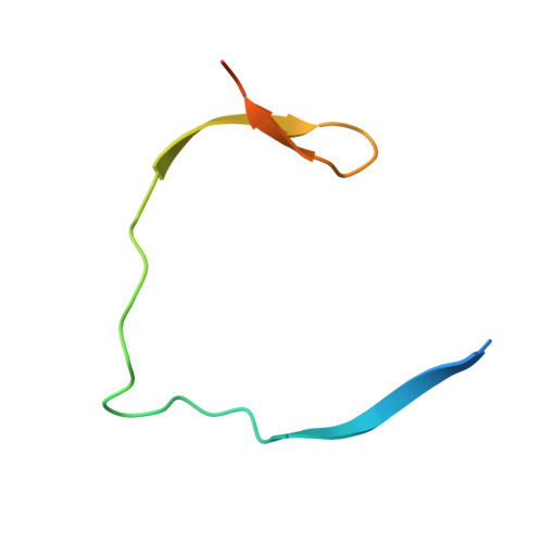 | |
UniProt | |||||
Find proteins for Q32ZE1 (Zika virus) Explore Q32ZE1 Go to UniProtKB: Q32ZE1 | |||||
Entity Groups | |||||
| Sequence Clusters | 30% Identity50% Identity70% Identity90% Identity95% Identity100% Identity | ||||
| UniProt Group | Q32ZE1 | ||||
Sequence AnnotationsExpand | |||||
| |||||
Entity ID: 2 | |||||
|---|---|---|---|---|---|
| Molecule | Chains | Sequence Length | Organism | Details | Image |
| Serine protease NS3 | 178 | Zika virus | Mutation(s): 0 EC: 3.4.21.91 (PDB Primary Data), 3.6.1.15 (PDB Primary Data), 3.6.4.13 (PDB Primary Data) | 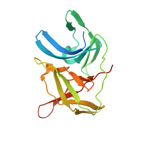 | |
UniProt | |||||
Find proteins for Q32ZE1 (Zika virus) Explore Q32ZE1 Go to UniProtKB: Q32ZE1 | |||||
Entity Groups | |||||
| Sequence Clusters | 30% Identity50% Identity70% Identity90% Identity95% Identity100% Identity | ||||
| UniProt Group | Q32ZE1 | ||||
Sequence AnnotationsExpand | |||||
| |||||
| Ligands 2 Unique | |||||
|---|---|---|---|---|---|
| ID | Chains | Name / Formula / InChI Key | 2D Diagram | 3D Interactions | |
| JW6 (Subject of Investigation/LOI) Query on JW6 | D [auth B] | 1-[(8R,15S,18S)-15-(4-azanylbutyl)-18-[(3-azanyl-4-oxidanyl-phenyl)methyl]-4,7,14,17,20-pentakis(oxidanylidene)-3,6,13,16,19-pentazabicyclo[20.3.1]hexacosa-1(25),22(26),23-trien-8-yl]guanidine C33 H48 N10 O6 RKZWERWMSWLYFI-NXCFDTQHSA-N |  | ||
| GOL Query on GOL | C [auth B] | GLYCEROL C3 H8 O3 PEDCQBHIVMGVHV-UHFFFAOYSA-N |  | ||
| Length ( Å ) | Angle ( ˚ ) |
|---|---|
| a = 48.918 | α = 90 |
| b = 60.253 | β = 90 |
| c = 82.385 | γ = 90 |
| Software Name | Purpose |
|---|---|
| PHENIX | refinement |
| XDS | data scaling |
| XDS | data reduction |
| PHASER | phasing |
| Coot | model building |
| Funding Organization | Location | Grant Number |
|---|---|---|
| Not funded | -- |
RCSB PDB (citation) is hosted by
RCSB PDB is a member of the

