7ZJB

Structural and functional characterization of the bacterial lytic polysaccharide Monooxygenase ScLPMO10D


Experimental Data Snapshot

  • Method: X-RAY DIFFRACTION
  • Resolution: 1.37 Å
  • R-Value Free: 0.152 
  • R-Value Work: 0.129 
  • R-Value Observed: 0.130 

wwPDB Validation   3D Report Full Report


This is version 1.1 of the entry. See complete history


Literature

Structural and functional characterization of the catalytic domain of a cell-wall anchored bacterial lytic polysaccharide monooxygenase from Streptomyces coelicolor.

Votvik, A.K.Rohr, A.K.Bissaro, B.Stepnov, A.A.Sorlie, M.Eijsink, V.G.H.Forsberg, Z.

(2023) Sci Rep 13: 5345-5345

  • DOI: https://doi.org/10.1038/s41598-023-32263-7
  • Primary Citation of Related Structures:  
    7ZJB

  • PubMed Abstract: 

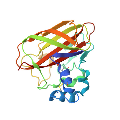
    Bacterial lytic polysaccharide monooxygenases (LPMOs) are known to oxidize the most abundant and recalcitrant polymers in Nature, namely cellulose and chitin. The genome of the model actinomycete Streptomyces coelicolor A3(2) encodes seven putative LPMOs, of which, upon phylogenetic analysis, four group with typical chitin-oxidizing LPMOs, two with typical cellulose-active LPMOs, and one which stands out by being part of a subclade of non-characterized enzymes. The latter enzyme, called ScLPMO10D, and most of the enzymes found in this subclade are unique, not only because of variation in the catalytic domain, but also as their C-terminus contains a cell wall sorting signal (CWSS), which flags the LPMO for covalent anchoring to the cell wall. Here, we have produced a truncated version of ScLPMO10D without the CWSS and determined its crystal structure, EPR spectrum, and various functional properties. While showing several structural and functional features typical for bacterial cellulose active LPMOs, ScLPMO10D is only active on chitin. Comparison with two known chitin-oxidizing LPMOs of different taxa revealed interesting functional differences related to copper reactivity. This study contributes to our understanding of the biological roles of LPMOs and provides a foundation for structural and functional comparison of phylogenetically distant LPMOs with similar substrate specificities.


  • Organizational Affiliation

    Faculty of Chemistry, Biotechnology, and Food Science, The Norwegian University of Life Sciences (NMBU), 1432, Ås, Norway.


Macromolecules
Find similar proteins by:  (by identity cutoff)  |  3D Structure
Entity ID: 1
MoleculeChains Sequence LengthOrganismDetailsImage
Putative secreted cellulose-binding proteinA [auth B]181Streptomyces coelicolor A3(2)Mutation(s): 0 
Gene Names: SCO1734
UniProt
Find proteins for Q9S296 (Streptomyces coelicolor (strain ATCC BAA-471 / A3(2) / M145))
Explore Q9S296 
Go to UniProtKB:  Q9S296
Entity Groups  
Sequence Clusters30% Identity50% Identity70% Identity90% Identity95% Identity100% Identity
UniProt GroupQ9S296
Sequence Annotations
Expand
  • Reference Sequence
Small Molecules
Ligands 3 Unique
IDChains Name / Formula / InChI Key2D Diagram3D Interactions
SO4
Query on SO4

Download Ideal Coordinates CCD File 
C [auth B]
D [auth B]
E [auth B]
F [auth B]
G [auth B]
C [auth B],
D [auth B],
E [auth B],
F [auth B],
G [auth B],
H [auth B],
I [auth B],
J [auth B],
K [auth B],
L [auth B]
SULFATE ION
O4 S
QAOWNCQODCNURD-UHFFFAOYSA-L
CU (Subject of Investigation/LOI)
Query on CU

Download Ideal Coordinates CCD File 
B
COPPER (II) ION
Cu
JPVYNHNXODAKFH-UHFFFAOYSA-N
NA
Query on NA

Download Ideal Coordinates CCD File 
M [auth B]SODIUM ION
Na
FKNQFGJONOIPTF-UHFFFAOYSA-N
Experimental Data & Validation

Experimental Data

  • Method: X-RAY DIFFRACTION
  • Resolution: 1.37 Å
  • R-Value Free: 0.152 
  • R-Value Work: 0.129 
  • R-Value Observed: 0.130 
  • Space Group: P 43 21 2
Unit Cell:
Length ( Å )Angle ( ˚ )
a = 59.08α = 90
b = 59.08β = 90
c = 145.718γ = 90
Software Package:
Software NamePurpose
REFMACrefinement
XDSdata reduction
pointlessdata scaling
PHASERphasing

Structure Validation

View Full Validation Report


Entry History & Funding Information

Deposition Data


Funding OrganizationLocationGrant Number
Novo Nordisk FoundationDenmarkNNF18OC0055736
Research Council of NorwayNorway301022
Research Council of NorwayNorway262853

Revision History  (Full details and data files)

  • Version 1.0: 2023-04-19
    Type: Initial release
  • Version 1.1: 2024-02-07
    Changes: Data collection, Refinement description