7ZJ7

X-31 Hemagglutinin Precursor HA0 at pH 4.8


Experimental Data Snapshot

  • Method: ELECTRON MICROSCOPY
  • Resolution: 3.95 Å
  • Aggregation State: PARTICLE 
  • Reconstruction Method: SINGLE PARTICLE 

wwPDB Validation   3D Report Full Report


This is version 1.0 of the entry. See complete history


Literature

Reversible structural changes in the influenza hemagglutinin precursor at membrane fusion pH.

Garcia-Moro, E.Zhang, J.Calder, L.J.Brown, N.R.Gamblin, S.J.Skehel, J.J.Rosenthal, P.B.

(2022) Proc Natl Acad Sci U S A 119: e2208011119-e2208011119

  • DOI: https://doi.org/10.1073/pnas.2208011119
  • Primary Citation of Related Structures:  
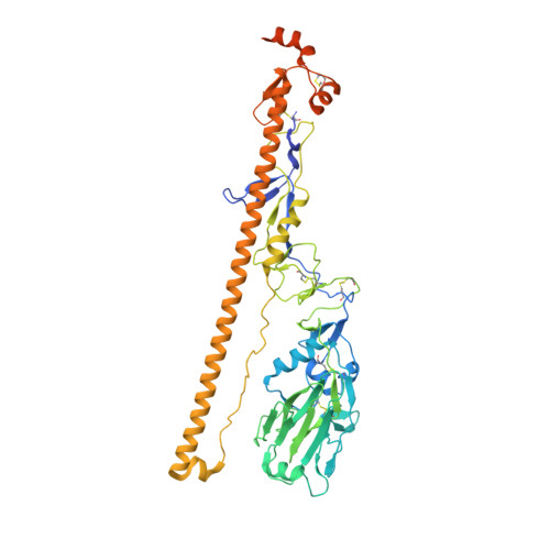
    7ZJ6, 7ZJ7, 7ZJ8

  • PubMed Abstract: 

    The subunits of the influenza hemagglutinin (HA) trimer are synthesized as single-chain precursors (HA0s) that are proteolytically cleaved into the disulfide-linked polypeptides HA1 and HA2. Cleavage is required for activation of membrane fusion at low pH, which occurs at the beginning of infection following transfer of cell-surface-bound viruses into endosomes. Activation results in extensive changes in the conformation of cleaved HA. To establish the overall contribution of cleavage to the mechanism of HA-mediated membrane fusion, we used cryogenic electron microscopy (cryo-EM) to directly image HA0 at neutral and low pH. We found extensive pH-induced structural changes, some of which were similar to those described for intermediates in the refolding of cleaved HA at low pH. They involve a partial extension of the long central coiled coil formed by melting of the preexisting secondary structure, threading it between the membrane-distal domains, and subsequent refolding as extended helices. The fusion peptide, covalently linked at its N terminus, adopts an amphipathic helical conformation over part of its length and is repositioned and packed against a complementary surface groove of conserved residues. Furthermore, and in contrast to cleaved HA, the changes in HA0 structure at low pH are reversible on reincubation at neutral pH. We discuss the implications of covalently restricted HA0 refolding for the cleaved HA conformational changes that mediate membrane fusion and for the action of antiviral drug candidates and cross-reactive anti-HA antibodies that can block influenza infectivity.


  • Organizational Affiliation

    Structural Biology of Cells and Viruses Laboratory, Francis Crick Institute, NW1 AT London, United Kingdom.


Macromolecules
Find similar proteins by:  (by identity cutoff)  |  3D Structure
Entity ID: 1
MoleculeChains Sequence LengthOrganismDetailsImage
Hemagglutinin,Fibritin
A, B, C
554Influenza A virus (A/Aichi/2/1968(H3N2))Tequatrovirus T4
This entity is chimeric
Mutation(s): 1 
Gene Names: HAwac
UniProt
Find proteins for P03437 (Influenza A virus (strain A/Aichi/2/1968 H3N2))
Explore P03437 
Go to UniProtKB:  P03437
Find proteins for P10104 (Enterobacteria phage T4)
Explore P10104 
Go to UniProtKB:  P10104
Entity Groups  
Sequence Clusters30% Identity50% Identity70% Identity90% Identity95% Identity100% Identity
UniProt GroupsP03437P10104
Sequence Annotations
Expand
  • Reference Sequence
Oligosaccharides

Help

Entity ID: 2
MoleculeChains Length2D Diagram Glycosylation3D Interactions
2-acetamido-2-deoxy-beta-D-glucopyranose-(1-4)-2-acetamido-2-deoxy-beta-D-glucopyranose
D, F, H, J, M
D, F, H, J, M, N
2N-Glycosylation
Glycosylation Resources
GlyTouCan:  G42666HT
GlyCosmos:  G42666HT
GlyGen:  G42666HT
Entity ID: 3
MoleculeChains Length2D Diagram Glycosylation3D Interactions
beta-D-mannopyranose-(1-4)-2-acetamido-2-deoxy-beta-D-glucopyranose-(1-4)-[alpha-L-fucopyranose-(1-6)]2-acetamido-2-deoxy-beta-D-glucopyranose
E, I, L
4N-Glycosylation
Glycosylation Resources
GlyTouCan:  G32152BH
GlyCosmos:  G32152BH
GlyGen:  G32152BH
Entity ID: 4
MoleculeChains Length2D Diagram Glycosylation3D Interactions
beta-D-mannopyranose-(1-4)-2-acetamido-2-deoxy-beta-D-glucopyranose-(1-4)-2-acetamido-2-deoxy-beta-D-glucopyranose
G, K, O
3N-Glycosylation
Glycosylation Resources
GlyTouCan:  G15407YE
GlyCosmos:  G15407YE
GlyGen:  G15407YE
Experimental Data & Validation

Experimental Data

  • Method: ELECTRON MICROSCOPY
  • Resolution: 3.95 Å
  • Aggregation State: PARTICLE 
  • Reconstruction Method: SINGLE PARTICLE 
EM Software:
TaskSoftware PackageVersion
RECONSTRUCTIONcryoSPARC3.1
MODEL REFINEMENTREFMAC5

Structure Validation

View Full Validation Report


Entry History & Funding Information

Deposition Data


Funding OrganizationLocationGrant Number
Wellcome TrustUnited Kingdom219946/Z/19/Z
The Francis Crick InstituteUnited Kingdom--

Revision History  (Full details and data files)

  • Version 1.0: 2022-08-17
    Type: Initial release