7YUY

Structure of a mutated membrane-bound glycosyltransferase


Experimental Data Snapshot

  • Method: ELECTRON MICROSCOPY
  • Resolution: 3.50 Å
  • Aggregation State: PARTICLE 
  • Reconstruction Method: SINGLE PARTICLE 

wwPDB Validation   3D Report Full Report


This is version 1.2 of the entry. See complete history


Literature

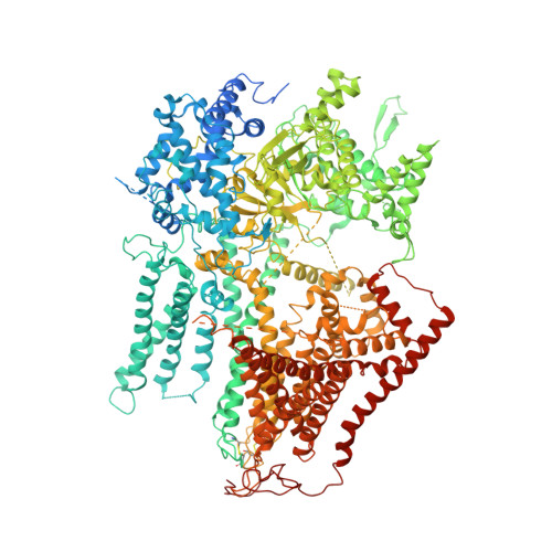
Structural and mechanistic insights into fungal beta-1,3-glucan synthase FKS1.

Hu, X.Yang, P.Chai, C.Liu, J.Sun, H.Wu, Y.Zhang, M.Zhang, M.Liu, X.Yu, H.

(2023) Nature 616: 190-198

  • DOI: https://doi.org/10.1038/s41586-023-05856-5
  • Primary Citation of Related Structures:  
    7XE4, 7YUY

  • PubMed Abstract: 

    The membrane-integrated synthase FKS is involved in the biosynthesis of β-1,3-glucan, the core component of the fungal cell wall 1,2 . FKS is the target of widely prescribed antifungal drugs, including echinocandin and ibrexafungerp 3,4 . Unfortunately, the mechanism of action of FKS remains enigmatic and this has hampered development of more effective medicines targeting the enzyme. Here we present the cryo-electron microscopy structures of Saccharomyces cerevisiae FKS1 and the echinocandin-resistant mutant FKS1(S643P). These structures reveal the active site of the enzyme at the membrane-cytoplasm interface and a glucan translocation path spanning the membrane bilayer. Multiple bound lipids and notable membrane distortions are observed in the FKS1 structures, suggesting active FKS1-membrane interactions. Echinocandin-resistant mutations are clustered at a region near TM5-6 and TM8 of FKS1. The structure of FKS1(S643P) reveals altered lipid arrangements in this region, suggesting a drug-resistant mechanism of the mutant enzyme. The structures, the catalytic mechanism and the molecular insights into drug-resistant mutations of FKS1 revealed in this study advance the mechanistic understanding of fungal β-1,3-glucan biosynthesis and establish a foundation for developing new antifungal drugs by targeting FKS.


  • Organizational Affiliation

    Department of Biochemistry and Molecular Biology, School of Basic Medicine, Tongji Medical College, Huazhong University of Science and Technology, Wuhan, China.


Macromolecules
Find similar proteins by:  (by identity cutoff)  |  3D Structure
Entity ID: 1
MoleculeChains Sequence LengthOrganismDetailsImage
1,3-beta-glucan synthase component FKS1A [auth F]1,876Saccharomyces cerevisiaeMutation(s): 1 
Gene Names: FKS1CND1CWH53ETG1GLS1GSC1PBR1YLR342WL8300.6
EC: 2.4.1.34
Membrane Entity: Yes 
UniProt
Find proteins for P38631 (Saccharomyces cerevisiae (strain ATCC 204508 / S288c))
Explore P38631 
Go to UniProtKB:  P38631
Entity Groups  
Sequence Clusters30% Identity50% Identity70% Identity90% Identity95% Identity100% Identity
UniProt GroupP38631
Sequence Annotations
Expand
  • Reference Sequence
Oligosaccharides

Help

Entity ID: 2
MoleculeChains Length2D Diagram Glycosylation3D Interactions
2-acetamido-2-deoxy-beta-D-glucopyranose-(1-4)-2-acetamido-2-deoxy-beta-D-glucopyranoseB [auth A]2N/A
Glycosylation Resources
GlyTouCan:  G42666HT
GlyCosmos:  G42666HT
GlyGen:  G42666HT
Entity ID: 3
MoleculeChains Length2D Diagram Glycosylation3D Interactions
beta-D-glucopyranose-(1-3)-beta-D-glucopyranose-(1-3)-beta-D-glucopyranose-(1-3)-beta-D-glucopyranose
C
4N/A
Glycosylation Resources
GlyTouCan:  G87841AX
GlyCosmos:  G87841AX
GlyGen:  G87841AX
Small Molecules
Ligands 6 Unique
IDChains Name / Formula / InChI Key2D Diagram3D Interactions
XKP
Query on XKP

Download Ideal Coordinates CCD File 
BA [auth F](11R,14S)-17-amino-14-hydroxy-8,14-dioxo-9,13,15-trioxa-14lambda~5~-phosphaheptadecan-11-yl decanoate
C23 H46 N O8 P
VYUSSAVVRCGYLY-OAQYLSRUSA-N
C14
Query on C14

Download Ideal Coordinates CCD File 
Q [auth F]TETRADECANE
C14 H30
BGHCVCJVXZWKCC-UHFFFAOYSA-N
D12
Query on D12

Download Ideal Coordinates CCD File 
AA [auth F],
Y [auth F],
Z [auth F]
DODECANE
C12 H26
SNRUBQQJIBEYMU-UHFFFAOYSA-N
D10
Query on D10

Download Ideal Coordinates CCD File 
K [auth F]
L [auth F]
M [auth F]
N [auth F]
O [auth F]
K [auth F],
L [auth F],
M [auth F],
N [auth F],
O [auth F],
P [auth F]
DECANE
C10 H22
DIOQZVSQGTUSAI-UHFFFAOYSA-N
DD9
Query on DD9

Download Ideal Coordinates CCD File 
D [auth F]
E [auth F]
F
G [auth F]
H [auth F]
D [auth F],
E [auth F],
F,
G [auth F],
H [auth F],
I [auth F],
J [auth F]
nonane
C9 H20
BKIMMITUMNQMOS-UHFFFAOYSA-N
HP6
Query on HP6

Download Ideal Coordinates CCD File 
R [auth F]
S [auth F]
T [auth F]
U [auth F]
V [auth F]
R [auth F],
S [auth F],
T [auth F],
U [auth F],
V [auth F],
W [auth F],
X [auth F]
HEPTANE
C7 H16
IMNFDUFMRHMDMM-UHFFFAOYSA-N
Experimental Data & Validation

Experimental Data

  • Method: ELECTRON MICROSCOPY
  • Resolution: 3.50 Å
  • Aggregation State: PARTICLE 
  • Reconstruction Method: SINGLE PARTICLE 

Structure Validation

View Full Validation Report


Entry History & Funding Information

Deposition Data


Funding OrganizationLocationGrant Number
National Natural Science Foundation of China (NSFC)China--

Revision History  (Full details and data files)

  • Version 1.0: 2023-03-29
    Type: Initial release
  • Version 1.1: 2023-04-05
    Changes: Database references
  • Version 1.2: 2023-04-19
    Changes: Database references