


Crystal structure of mango alpha1,3/alpha1,4-fucosyltransferase elucidates unique elements strictly regulating its type I-dominant acceptor preference
Okada, T., Teramoto, T., Ihara, H., Ikeda, Y., Kakuta, Y.To be published.
Experimental Data Snapshot
Entity ID: 1 | |||||
|---|---|---|---|---|---|
| Molecule | Chains | Sequence Length | Organism | Details | Image |
| Fucosyltransferase | 410 | Mangifera indica | Mutation(s): 0 Gene Names: FUT13 EC: 2.4.1 | 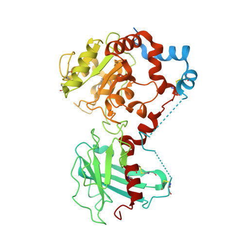 | |
UniProt | |||||
Find proteins for A0A292GKJ7 (Mangifera indica) Explore A0A292GKJ7 Go to UniProtKB: A0A292GKJ7 | |||||
Entity Groups | |||||
| Sequence Clusters | 30% Identity50% Identity70% Identity90% Identity95% Identity100% Identity | ||||
| UniProt Group | A0A292GKJ7 | ||||
Sequence AnnotationsExpand | |||||
| |||||
| Ligands 5 Unique | |||||
|---|---|---|---|---|---|
| ID | Chains | Name / Formula / InChI Key | 2D Diagram | 3D Interactions | |
| NAG (Subject of Investigation/LOI) Query on NAG | AA [auth D], GA [auth E], M [auth B], PA [auth F], R [auth C] | 2-acetamido-2-deoxy-beta-D-glucopyranose C8 H15 N O6 OVRNDRQMDRJTHS-FMDGEEDCSA-N |  | ||
| PPV (Subject of Investigation/LOI) Query on PPV | FA [auth E] L [auth B] LA [auth F] Q [auth C] RA [auth G] | PYROPHOSPHATE H4 O7 P2 XPPKVPWEQAFLFU-UHFFFAOYSA-N |  | ||
| PO4 (Subject of Investigation/LOI) Query on PO4 | K [auth A] | PHOSPHATE ION O4 P NBIIXXVUZAFLBC-UHFFFAOYSA-K |  | ||
| GOL (Subject of Investigation/LOI) Query on GOL | BA [auth D] CA [auth D] DA [auth D] EA [auth D] HA [auth E] | GLYCEROL C3 H8 O3 PEDCQBHIVMGVHV-UHFFFAOYSA-N |  | ||
| ACY (Subject of Investigation/LOI) Query on ACY | IA [auth E] J [auth A] P [auth B] QA [auth F] TA [auth G] | ACETIC ACID C2 H4 O2 QTBSBXVTEAMEQO-UHFFFAOYSA-N |  | ||
| Length ( Å ) | Angle ( ˚ ) |
|---|---|
| a = 86.7 | α = 90 |
| b = 163.61 | β = 90 |
| c = 274.9 | γ = 90 |
| Software Name | Purpose |
|---|---|
| PHENIX | refinement |
| XDS | data reduction |
| XDS | data scaling |
| PHASER | phasing |
| Funding Organization | Location | Grant Number |
|---|---|---|
| Japan Society for the Promotion of Science (JSPS) | Japan | 20K06019 |
RCSB PDB (citation) is hosted by
RCSB PDB is a member of the

