


The crystal structure of AhpD from Pseudomonas aeruginosa
Xu, B.To be published.
Experimental Data Snapshot
wwPDB Validation 3D Report Full Report

Entity ID: 1 | |||||
|---|---|---|---|---|---|
| Molecule | Chains | Sequence Length | Organism | Details | Image |
| Carboxymuconolactone decarboxylase family protein | 145 | Pseudomonas aeruginosa | Mutation(s): 0 | 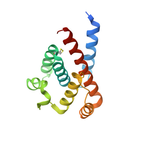 | |
UniProt | |||||
Find proteins for A0A2U2XNT2 (Pseudomonas aeruginosa) Explore A0A2U2XNT2 Go to UniProtKB: A0A2U2XNT2 | |||||
Entity Groups | |||||
| Sequence Clusters | 30% Identity50% Identity70% Identity90% Identity95% Identity100% Identity | ||||
| UniProt Group | A0A2U2XNT2 | ||||
Sequence AnnotationsExpand | |||||
| |||||
| Length ( Å ) | Angle ( ˚ ) |
|---|---|
| a = 92.976 | α = 90 |
| b = 92.976 | β = 90 |
| c = 65.957 | γ = 120 |
| Software Name | Purpose |
|---|---|
| Aimless | data scaling |
| PHENIX | refinement |
| PDB_EXTRACT | data extraction |
| XDS | data reduction |
| PHASER | phasing |
| Funding Organization | Location | Grant Number |
|---|---|---|
| Not funded | -- |
RCSB PDB (citation) is hosted by
RCSB PDB is a member of the

