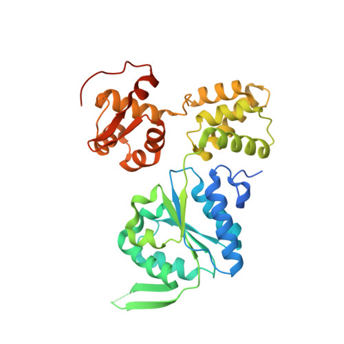
7X5B

Crystal structure of RuvB


Experimental Data Snapshot

  • Method: X-RAY DIFFRACTION
  • Resolution: 2.16 Å
  • R-Value Free: 0.217 
  • R-Value Work: 0.173 
  • R-Value Observed: 0.177 

wwPDB Validation   3D Report Full Report


Ligand Structure Quality Assessment 


This is version 1.2 of the entry. See complete history


Literature

Cryo-EM structure of the RuvAB-Holliday junction intermediate complex from Pseudomonas aeruginosa.

Zhang, X.Zhou, Z.Dai, L.Chao, Y.Liu, Z.Huang, M.Qu, Q.Lin, Z.

(2023) Front Plant Sci 14: 1139106-1139106

  • DOI: https://doi.org/10.3389/fpls.2023.1139106
  • Primary Citation of Related Structures:  
    7X5A, 7X5B, 7X7P, 7X7Q

  • PubMed Abstract: 

    Holliday junction (HJ) is a four-way structured DNA intermediate in homologous recombination. In bacteria, the HJ-specific binding protein RuvA and the motor protein RuvB together form the RuvAB complex to catalyze HJ branch migration. Pseudomonas aeruginosa ( P. aeruginosa , Pa) is a ubiquitous opportunistic bacterial pathogen that can cause serious infection in a variety of host species, including vertebrate animals, insects and plants. Here, we describe the cryo-Electron Microscopy (cryo-EM) structure of the RuvAB-HJ intermediate complex from P. aeruginosa . The structure shows that two RuvA tetramers sandwich HJ at the junction center and disrupt base pairs at the branch points of RuvB-free HJ arms. Eight RuvB subunits are recruited by the RuvA octameric core and form two open-rings to encircle two opposite HJ arms. Each RuvB subunit individually binds a RuvA domain III. The four RuvB subunits within the ring display distinct subdomain conformations, and two of them engage the central DNA duplex at both strands with their C-terminal β-hairpins. Together with the biochemical analyses, our structure implicates a potential mechanism of RuvB motor assembly onto HJ DNA.


  • Organizational Affiliation

    College of Chemistry, Fuzhou University, Fuzhou, China.


Macromolecules
Find similar proteins by:  (by identity cutoff)  |  3D Structure
Entity ID: 1
MoleculeChains Sequence LengthOrganismDetailsImage
Holliday junction ATP-dependent DNA helicase RuvB352Pseudomonas aeruginosa PAO1Mutation(s): 1 
Gene Names: ruvB
EC: 3.6.4.12
UniProt
Find proteins for Q51426 (Pseudomonas aeruginosa (strain ATCC 15692 / DSM 22644 / CIP 104116 / JCM 14847 / LMG 12228 / 1C / PRS 101 / PAO1))
Explore Q51426 
Go to UniProtKB:  Q51426
Entity Groups  
Sequence Clusters30% Identity50% Identity70% Identity90% Identity95% Identity100% Identity
UniProt GroupQ51426
Sequence Annotations
Expand
  • Reference Sequence
Small Molecules
Ligands 1 Unique
IDChains Name / Formula / InChI Key2D Diagram3D Interactions
ADP (Subject of Investigation/LOI)
Query on ADP

Download Ideal Coordinates CCD File 
B [auth A]ADENOSINE-5'-DIPHOSPHATE
C10 H15 N5 O10 P2
XTWYTFMLZFPYCI-KQYNXXCUSA-N
Experimental Data & Validation

Experimental Data

  • Method: X-RAY DIFFRACTION
  • Resolution: 2.16 Å
  • R-Value Free: 0.217 
  • R-Value Work: 0.173 
  • R-Value Observed: 0.177 
  • Space Group: P 65
Unit Cell:
Length ( Å )Angle ( ˚ )
a = 85.677α = 90
b = 85.677β = 90
c = 76.973γ = 120
Software Package:
Software NamePurpose
HKL-3000data reduction
HKL-3000data scaling
PHENIXrefinement
MOLREPphasing

Structure Validation

View Full Validation Report


Ligand Structure Quality Assessment 


Entry History & Funding Information

Deposition Data


Funding OrganizationLocationGrant Number
National Natural Science Foundation of China (NSFC)China31971222
National Natural Science Foundation of China (NSFC)China32171194

Revision History  (Full details and data files)

  • Version 1.0: 2023-03-08
    Type: Initial release
  • Version 1.1: 2023-09-20
    Changes: Data collection, Database references
  • Version 1.2: 2023-11-29
    Changes: Refinement description