


Crystal structure of multidomain beta-1,3(4)-glucanase
Jiang, Z.Q., Ma, J.W.To be published.
Experimental Data Snapshot
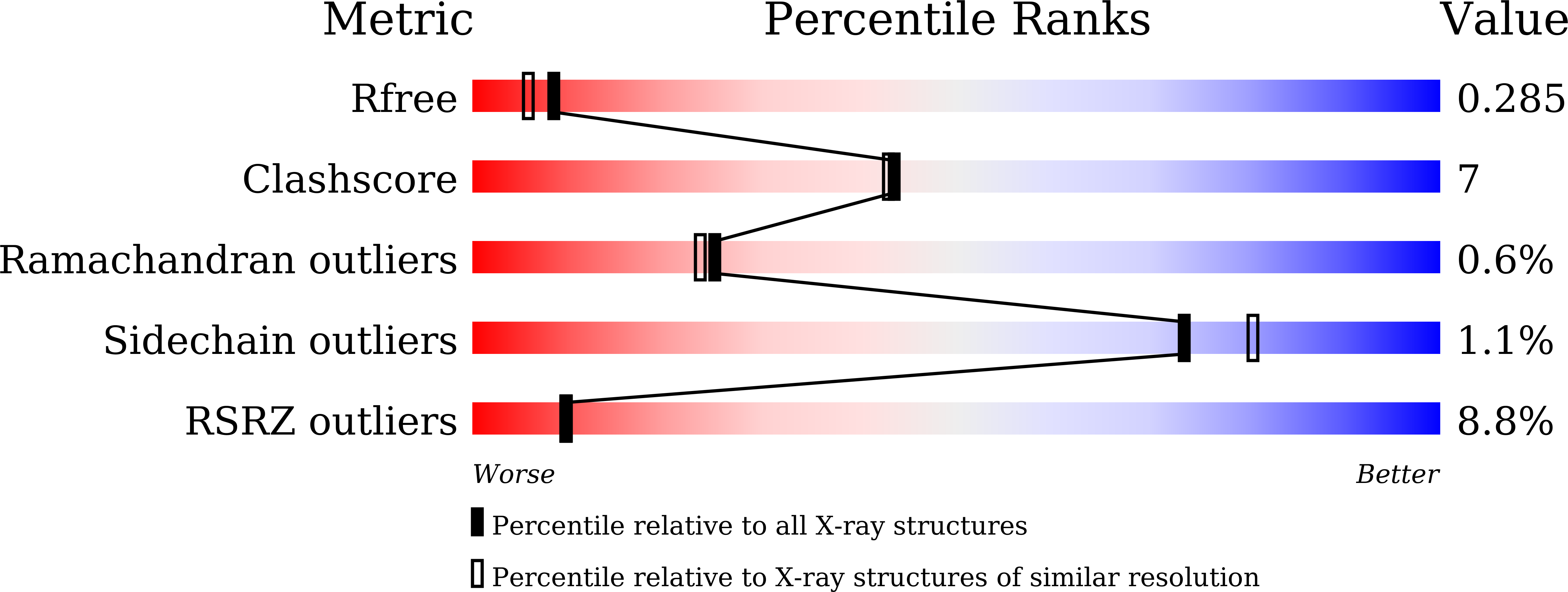
wwPDB Validation 3D Report Full Report
Entity ID: 1 | |||||
|---|---|---|---|---|---|
| Molecule | Chains | Sequence Length | Organism | Details | Image |
| beta-1,3(4)-glucanase | 480 | Pedobacter sp. | Mutation(s): 0 | 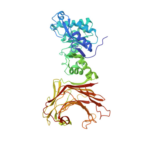 | |
UniProt | |||||
Find proteins for A0A1G7XNR7 (Pedobacter terrae) Explore A0A1G7XNR7 Go to UniProtKB: A0A1G7XNR7 | |||||
Entity Groups | |||||
| Sequence Clusters | 30% Identity50% Identity70% Identity90% Identity95% Identity100% Identity | ||||
| UniProt Group | A0A1G7XNR7 | ||||
Sequence AnnotationsExpand | |||||
| |||||
| Ligands 2 Unique | |||||
|---|---|---|---|---|---|
| ID | Chains | Name / Formula / InChI Key | 2D Diagram | 3D Interactions | |
| GOL Query on GOL | D [auth A], E [auth A] | GLYCEROL C3 H8 O3 PEDCQBHIVMGVHV-UHFFFAOYSA-N |  | ||
| CA Query on CA | B [auth A], C [auth A] | CALCIUM ION Ca BHPQYMZQTOCNFJ-UHFFFAOYSA-N |  | ||
| Length ( Å ) | Angle ( ˚ ) |
|---|---|
| a = 120.94 | α = 90 |
| b = 120.94 | β = 90 |
| c = 70.05 | γ = 120 |
| Software Name | Purpose |
|---|---|
| HKL-3000 | data collection |
| HKL-3000 | data scaling |
| PHENIX | model building |
| PHENIX | refinement |
| Funding Organization | Location | Grant Number |
|---|---|---|
| National Natural Science Foundation of China (NSFC) | China | -- |
RCSB PDB (citation) is hosted by
RCSB PDB is a member of the

