


Crystal structure of metagenomic beta-glycosidase MeBglD2 in complex with various saccharides
Matsuzawa, T., Watanabe, M., Nakamichi, Y., Akita, H., Yaoi, K.(2022) Appl Microbiol Biotechnol 106: 4539-4551
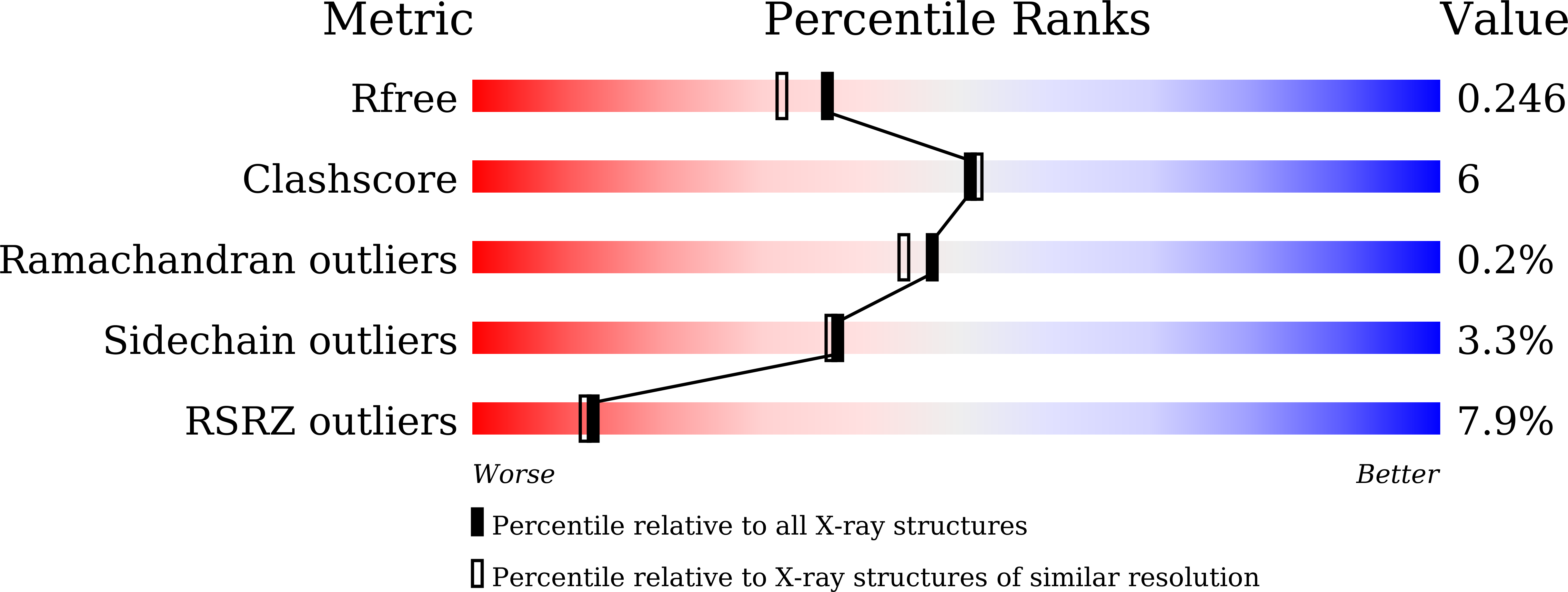
Experimental Data Snapshot
(2022) Appl Microbiol Biotechnol 106: 4539-4551
Entity ID: 1 | |||||
|---|---|---|---|---|---|
| Molecule | Chains | Sequence Length | Organism | Details | Image |
| Beta-glucosidase | 458 | metagenome | Mutation(s): 1 EC: 3.2.1.21 | 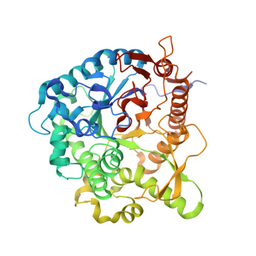 | |
UniProt | |||||
Find proteins for A0A1E1FFN6 (uncultured microorganism) Explore A0A1E1FFN6 Go to UniProtKB: A0A1E1FFN6 | |||||
Entity Groups | |||||
| Sequence Clusters | 30% Identity50% Identity70% Identity90% Identity95% Identity100% Identity | ||||
| UniProt Group | A0A1E1FFN6 | ||||
Sequence AnnotationsExpand | |||||
| |||||
| Ligands 2 Unique | |||||
|---|---|---|---|---|---|
| ID | Chains | Name / Formula / InChI Key | 2D Diagram | 3D Interactions | |
| PNW (Subject of Investigation/LOI) Query on PNW | B [auth A], C [auth A] | 4-nitrophenyl beta-D-glucopyranoside C12 H15 N O8 IFBHRQDFSNCLOZ-RMPHRYRLSA-N |  | ||
| SO4 (Subject of Investigation/LOI) Query on SO4 | D [auth A], E [auth A], F [auth A] | SULFATE ION O4 S QAOWNCQODCNURD-UHFFFAOYSA-L |  | ||
| Length ( Å ) | Angle ( ˚ ) |
|---|---|
| a = 201.847 | α = 90 |
| b = 201.847 | β = 90 |
| c = 201.847 | γ = 90 |
| Software Name | Purpose |
|---|---|
| REFMAC | refinement |
| DENZO | data reduction |
| SCALEPACK | data scaling |
| PHASER | phasing |
| Funding Organization | Location | Grant Number |
|---|---|---|
| Japan Society for the Promotion of Science (JSPS) | Japan | -- |
RCSB PDB (citation) is hosted by
RCSB PDB is a member of the

