7WDK

The structure of PldA-PA3488 complex


Experimental Data Snapshot

  • Method: ELECTRON MICROSCOPY
  • Resolution: 3.05 Å
  • Aggregation State: PARTICLE 
  • Reconstruction Method: SINGLE PARTICLE 

wwPDB Validation   3D Report Full Report


This is version 1.1 of the entry. See complete history


Literature

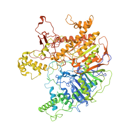
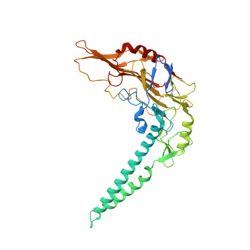
Structural insights into PA3488-mediated inactivation of Pseudomonas aeruginosa PldA

Yang, X.Li, Z.Zhao, L.She, Z.Gao, Z.Sui, S.F.Dong, Y.Li, Y.

(2022) Nat Commun 13: 5979-5979

  • DOI: https://doi.org/10.1038/s41467-022-33690-2
  • Primary Citation of Related Structures:  
    7V53, 7V55, 7WDK

  • PubMed Abstract: 

    PldA, a phospholipase D (PLD) effector, catalyzes hydrolysis of the phosphodiester bonds of glycerophospholipids-the main component of cell membranes-and assists the invasion of the opportunistic pathogen Pseudomonas aeruginosa. As a cognate immunity protein, PA3488 can inhibit the activity of PldA to avoid self-toxicity. However, the precise inhibitory mechanism remains elusive. We determine the crystal structures of full-length and truncated PldA and the cryogenic electron microscopy structure of the PldA-PA3488 complex. Structural analysis reveals that there are different intermediates of PldA between the "open" and "closed" states of the catalytic pocket, accompanied by significant conformational changes in the "lid" region and the peripheral helical domain. Through structure-based mutational analysis, we identify the key residues responsible for the enzymatic activity of PldA. Together, these data provide an insight into the molecular mechanisms of PldA invasion and its neutralization by PA3488, aiding future design of PLD-targeted inhibitors and drugs.


  • Organizational Affiliation

    Department of Biology, Southern University of Science and Technology, Shenzhen, 518055, Guangdong Province, China.


Macromolecules
Find similar proteins by:  (by identity cutoff)  |  3D Structure
Entity ID: 1
MoleculeChains Sequence LengthOrganismDetailsImage
Phospholipase D1,099Pseudomonas aeruginosa PAO1Mutation(s): 0 
Gene Names: pldAPA3487
UniProt
Find proteins for Q9HYC2 (Pseudomonas aeruginosa (strain ATCC 15692 / DSM 22644 / CIP 104116 / JCM 14847 / LMG 12228 / 1C / PRS 101 / PAO1))
Explore Q9HYC2 
Go to UniProtKB:  Q9HYC2
Entity Groups  
Sequence Clusters30% Identity50% Identity70% Identity90% Identity95% Identity100% Identity
UniProt GroupQ9HYC2
Sequence Annotations
Expand
  • Reference Sequence
Find similar proteins by:  (by identity cutoff)  |  3D Structure
Entity ID: 2
MoleculeChains Sequence LengthOrganismDetailsImage
Tli4_C domain-containing protein376Pseudomonas aeruginosaMutation(s): 0 
Gene Names: CGU42_05350E4V10_17995ECC04_007070GNQ48_30755
UniProt
Find proteins for A0A232D7B1 (Pseudomonas aeruginosa)
Explore A0A232D7B1 
Go to UniProtKB:  A0A232D7B1
Entity Groups  
Sequence Clusters30% Identity50% Identity70% Identity90% Identity95% Identity100% Identity
UniProt GroupA0A232D7B1
Sequence Annotations
Expand
  • Reference Sequence
Experimental Data & Validation

Experimental Data

  • Method: ELECTRON MICROSCOPY
  • Resolution: 3.05 Å
  • Aggregation State: PARTICLE 
  • Reconstruction Method: SINGLE PARTICLE 

Structure Validation

View Full Validation Report


Entry History & Funding Information

Deposition Data


Funding OrganizationLocationGrant Number
Chinese Academy of SciencesChina--

Revision History  (Full details and data files)

  • Version 1.0: 2022-10-26
    Type: Initial release
  • Version 1.1: 2022-11-09
    Changes: Database references