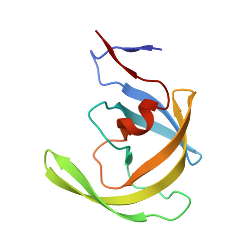
7WBS

Crystal structure of HIV-1 protease in complex with lactam derivative 2


Experimental Data Snapshot

  • Method: X-RAY DIFFRACTION
  • Resolution: 1.85 Å
  • R-Value Free: 0.200 
  • R-Value Work: 0.184 
  • R-Value Observed: 0.184 

wwPDB Validation   3D Report Full Report


Ligand Structure Quality Assessment 


This is version 1.1 of the entry. See complete history


Literature

Pocket-to-Lead: Structure-Based De Novo Design of Novel Non-peptidic HIV-1 Protease Inhibitors Using the Ligand Binding Pocket as a Template.

Kojima, E.Iimuro, A.Nakajima, M.Kinuta, H.Asada, N.Sako, Y.Nakata, Z.Uemura, K.Arita, S.Miki, S.Wakasa-Morimoto, C.Tachibana, Y.

(2022) J Med Chem 65: 6157-6170

  • DOI: https://doi.org/10.1021/acs.jmedchem.1c02217
  • Primary Citation of Related Structures:  
    7WBS, 7WCQ

  • PubMed Abstract: 

    A novel strategy for lead identification that we have dubbed the "Pocket-to-Lead" strategy is demonstrated using HIV-1 protease as a model target. Sometimes, it is difficult to obtain hit compounds because of the difficulties in satisfying the complex pharmacophoric features. In this study, a virtual fragment hit which does not match all of the pharmacophore features but has key interactions and vectors that could grow into remaining pharmacophore features was optimized in silico . The designed compound 9 demonstrated weak but evident inhibitory activity (IC 50 = 54 μM), and the design concept was proven by the co-crystal structure. Then, structure-based drug design promptly gave compound 14 (IC 50 = 0.0071 μM, EC 50 = 0.86 μM), an almost 10,000-fold improvement in activity from 9 . The structure of the designed molecules proved to be novel with high synthetic feasibility, indicating the usefulness of this strategy to tackle tough targets with complex pharmacophore.


  • Organizational Affiliation

    Shionogi Pharmaceutical Research Center, 3-1-1 Futaba-cho, Toyonaka, Osaka 561-0825, Japan.


Macromolecules
Find similar proteins by:  (by identity cutoff)  |  3D Structure
Entity ID: 1
MoleculeChains Sequence LengthOrganismDetailsImage
Protease
A, B
99Human immunodeficiency virus 1Mutation(s): 0 
UniProt
Find proteins for P12497 (Human immunodeficiency virus type 1 group M subtype B (isolate NY5))
Explore P12497 
Go to UniProtKB:  P12497
Entity Groups  
Sequence Clusters30% Identity50% Identity70% Identity90% Identity95% Identity100% Identity
UniProt GroupP12497
Sequence Annotations
Expand
  • Reference Sequence
Small Molecules
Ligands 2 Unique
IDChains Name / Formula / InChI Key2D Diagram3D Interactions
8LT (Subject of Investigation/LOI)
Query on 8LT

Download Ideal Coordinates CCD File 
D [auth B](3~{R},4~{R})-1-[(4-methoxyphenyl)methyl]-3-(3-methylbutyl)-3-[4-methylsulfonyl-2-[(2~{S})-1-oxidanylpropan-2-yl]oxy-phenyl]-4-oxidanyl-pyrrolidin-2-one
C27 H37 N O7 S
QQVYMLIPGZJSRF-YDOVEJIGSA-N
GOL
Query on GOL

Download Ideal Coordinates CCD File 
C [auth A],
E [auth B]
GLYCEROL
C3 H8 O3
PEDCQBHIVMGVHV-UHFFFAOYSA-N
Experimental Data & Validation

Experimental Data

  • Method: X-RAY DIFFRACTION
  • Resolution: 1.85 Å
  • R-Value Free: 0.200 
  • R-Value Work: 0.184 
  • R-Value Observed: 0.184 
  • Space Group: P 21 21 2
Unit Cell:
Length ( Å )Angle ( ˚ )
a = 58.254α = 90
b = 85.952β = 90
c = 46.62γ = 90
Software Package:
Software NamePurpose
REFMACrefinement
HKL-2000data reduction
HKL-2000data scaling
MOLREPphasing
PDB_EXTRACTdata extraction

Structure Validation

View Full Validation Report


Ligand Structure Quality Assessment 


Entry History & Funding Information

Deposition Data


Funding OrganizationLocationGrant Number
Not funded--

Revision History  (Full details and data files)

  • Version 1.0: 2022-11-02
    Type: Initial release
  • Version 1.1: 2023-11-29
    Changes: Data collection, Refinement description